Fat has acquired a poor reputation.
It is frequently described as something to burn, lose, remove or avoid. Excessive accumulation of certain types of fat is undoubtedly associated with metabolic disease. However, this does not mean that all fat is unhealthy—or even that all fat behaves in the same way.
Fat is not one uniform substance distributed randomly around the body. Adipose tissue is a living, vascular, innervated and highly specialised connective tissue. Its structure and function change according to where it is located.
The fat surrounding the abdominal organs behaves partly like an endocrine organ, communicating chemically with the rest of the body. By contrast, the fat beneath the heel is arranged as an intricate load-bearing structure that protects the heel bone from the repeated forces of standing, walking and running.
One fat depot can influence blood glucose and systemic inflammation. Another can act like a biological shock-absorbing system.
Understanding this difference helps us appreciate that healthy fat is not simply excess material. In some locations, it is an essential anatomical organ.
There is no single type of “body fat”
The word fat usually refers to adipose tissue, but adipose tissue exists in several anatomically and biologically distinct depots.
These include:
- visceral fat surrounding the abdominal organs;
- abdominal subcutaneous fat beneath the skin;
- fat around the kidneys, heart and blood vessels;
- bone marrow fat;
- fat within and around joints;
- the cushioning fat pads of the palms and soles;
- and specialised fat beneath the heel and forefoot.
Although these tissues contain adipocytes—cells capable of storing lipid—the surrounding connective tissue, blood supply, immune-cell population, cellular signalling and mechanical environment differ considerably.
This is why the location of fat can be as important as the total amount.
Studies using computed tomography and magnetic resonance imaging have shown that visceral abdominal fat generally has a stronger relationship with insulin resistance and adverse cardiometabolic markers than abdominal subcutaneous fat. Even this distinction is not absolute: subcutaneous adipose tissue can also become dysfunctional, while the capacity to store energy safely in subcutaneous tissue may help prevent lipid from accumulating in the liver, muscles and other organs (Preis et al., 2010; Neeland et al., 2013).
Is abdominal fat really an organ?
Adipose tissue was once regarded primarily as a passive energy-storage compartment. It is now recognised as an endocrine and immune-active tissue.
Adipocytes and the other cells living within adipose tissue produce chemical messengers called adipokines. These include leptin, adiponectin, chemerin, resistin and many other signalling proteins. Adipose tissue can also contribute to the production of inflammatory mediators, coagulation-related factors and proteins involved in vascular and glucose regulation.
Some of these signals are beneficial. Adiponectin, for example, is generally associated with healthier glucose and lipid metabolism. Others may become harmful when an adipose depot enlarges, becomes hypoxic, attracts inflammatory immune cells or develops abnormal free-fatty-acid release.
Therefore, visceral fat does not cause metabolic syndrome simply because it is fat. The problem is more accurately described as dysfunctional adipose tissue.
Human tissue studies demonstrate that visceral and subcutaneous abdominal fat do not release identical combinations of signalling molecules. In one paired-biopsy study, subcutaneous tissue released more adiponectin, while visceral tissue released more chemerin and dipeptidyl peptidase-4. This illustrates that the anatomical location of an adipose depot can alter its biological behaviour (Svensson et al., 2014).
Visceral fat also drains partly through the portal circulation towards the liver. Consequently, increased free-fatty-acid and inflammatory signalling from this depot may have a particularly direct effect on hepatic glucose and lipid metabolism.
Is heel fat completely different from abdominal fat?
At the level of the individual cell, heel adipocytes still resemble adipocytes elsewhere in the body. The heel fat pad is not made from an entirely different substance.
The crucial difference lies in how those cells are organised.
The heel contains a relatively small amount of adipose tissue enclosed within a highly reinforced system of collagenous and elastic partitions. Its clinically dominant purpose is therefore not whole-body energy storage or systemic hormone production. Its principal role is mechanical protection.
The heel fat pad is nevertheless alive. Anatomical studies have demonstrated a significant blood supply, free nerve endings and pressure-sensitive structures, including Pacinian corpuscles, within the plantar fat pads (Jahss et al., 1992).
It would consequently be inaccurate to call the heel fat pad metabolically inert. However, because it is small and structurally compartmentalised, there is no convincing evidence that a healthy heel fat pad makes a meaningful contribution to metabolic syndrome.
Its primary influence is local rather than systemic.
The anatomy of the heel fat pad
The heel fat pad sits between the plantar skin and the inferior surface of the calcaneus—the heel bone.
In an adult, the unloaded pad is commonly approximately one to two centimetres thick, although thickness varies with body size, age, sex, measurement method and loading conditions. Thickness alone does not tell us whether the pad is healthy.
The pad is traditionally described as having two interconnected regions.
The superficial microchamber layer
Immediately beneath the thick plantar skin lies a layer containing numerous relatively small fat compartments called microchambers.
These chambers are tightly packed and supported by fine fibrous septa. They appear to deform relatively little under normal loading.
The microchamber layer may help:
- stabilise the plantar skin;
- resist shear;
- prevent excessive sideways movement of the deeper fat;
- protect the skin from localised pressure;
- and maintain a durable interface between the ground and the foot.
The deeper macrochamber layer
Closer to the calcaneus is the macrochamber layer, which contains larger compartments.
These larger chambers undergo considerably more deformation as body weight is transferred onto the heel. They are therefore considered the principal compressible component of the heel pad.
During loading, the macrochambers shorten vertically and expand in other directions. Their expansion is controlled by strong fibrous walls and by attachments between the plantar skin, fascia and calcaneus.
Recent ultrasound studies support the concept that the superficial and deep layers behave differently. The macrochamber layer shows greater load-dependent change, while the microchamber layer remains comparatively stable (Maemichi et al., 2024a; Maemichi et al., 2024b).
A collection of miniature hydraulic cushions
The heel pad can be imagined as a collection of fluid-filled or gel-like compartments held inside a reinforced mesh.
This is not literally a bag of liquid. Instead, each chamber contains lipid-rich adipocytes, connective tissue, extracellular fluid, blood vessels and nerves. Because biological tissues contain large amounts of water and are difficult to compress volumetrically, pressure applied from above causes the tissue to change shape rather than simply collapse.
The fibrous septa limit how far the fat can move.
This closed-cell arrangement allows each chamber to act like a miniature hydrostatic system. The chambers distribute load to adjoining compartments while the collagen and elastic fibres resist excessive spreading.
Histological studies of atrophied heel pads have found fragmented and widened septal walls, demonstrating that failure of the supporting framework may be at least as important as a reduction in the quantity of fat itself (Buschmann et al., 1995).
How does the heel fat pad absorb shock?
Calling the heel fat pad a cushion is correct, but incomplete.
A household cushion is usually soft throughout its compression. The heel fat pad is more sophisticated. It demonstrates nonlinear viscoelastic behaviour.
Nonlinear behaviour
At the beginning of heel loading, the pad is relatively compliant. This allows it to deform and spread load without producing an abrupt pressure peak.
As compression increases, the pad progressively stiffens. The collagenous septa become tensioned, the fat compartments become increasingly constrained and further deformation becomes more difficult.
This prevents the heel from simply “bottoming out” against the calcaneus.
The result is a tissue that is:
- soft enough initially to accommodate impact;
- progressively firmer as compression increases;
- and highly resistant near the end of its safe deformation range.
Viscoelastic behaviour
The heel pad is also viscoelastic. Its response depends partly on how quickly it is loaded and on its recent loading history.
When the heel is compressed, some mechanical energy is temporarily stored and some is dissipated as heat. The loading and unloading curves are therefore not identical—a phenomenon called hysteresis.
This energy dissipation helps reduce rapid loading transients transmitted towards the calcaneus and lower limb.
Experimental studies show that heel-pad material properties change with strain rate, meaning the tissue responds differently to slow standing pressure, walking, running and sudden impact (Grigoriadis et al., 2017).
Does the heel fat pad completely absorb the force of running?
No.
The heel pad is an important local shock attenuator, but it cannot eliminate the forces of locomotion. Impact management is shared between:
- the heel pad;
- the shoe and ground surface;
- ankle and foot motion;
- the plantar fascia;
- the Achilles tendon;
- muscle activity;
- knee and hip flexion;
- and movement of the rest of the body.
In an in-vivo cineradiographic study, barefoot running compressed the heel pad considerably more than shod running. Barefoot deformation approached approximately 60% in the small sample, compared with approximately 36% when shoes were worn. This suggests that footwear can reduce how close the biological pad comes to its compression limit (De Clercq, Aerts and Kunnen, 1994).
During ordinary walking, the heel pad may deform by roughly one centimetre. Research has suggested that it can operate surprisingly close to its physiological deformation range even at walking speeds (Wearing et al., 2009).
This does not mean walking is damaging. Healthy tissues are designed to experience load. It means that the pad performs substantial mechanical work during every step.
What makes the heel fat pad special?
Several features distinguish it from ordinary subcutaneous abdominal fat.
1. It is securely anchored
Abdominal subcutaneous fat is comparatively mobile beneath the skin. The heel pad is bound by fibrous retinacula extending between the skin, deeper fascia and calcaneal region.
These attachments help prevent the pad from being squeezed completely away from the area that requires protection.
2. It has separate functional layers
Its superficial microchambers and deep macrochambers appear to perform complementary roles. One contributes more to skin stability and shear control; the other contributes more to compression and energy dissipation.
3. It becomes stiffer as it is compressed
This protects it against complete collapse and reduces the risk of the calcaneus striking through the soft tissue.
4. It must resist both compression and shear
The heel is not loaded vertically alone. At heel contact, braking forces and foot motion produce horizontal shear. The fat pad must therefore deform in three dimensions while remaining centred beneath the calcaneus.
5. It contains sensory and vascular structures
The heel pad contributes to the sensory interface between the foot and the ground. Its blood vessels and nerves also mean that damage, fibrosis, oedema or excessive compression may become painful.
6. Its external shape is anatomically organised
Three-dimensional imaging has shown that the heel pad has a consistent, complex geometry rather than being an unstructured mass of fat. Its contours appear adapted to the form of the calcaneus and the areas exposed to plantar loading (Campanelli et al., 2011).
How long does a heel fat pad last?
There is no fixed expiry date.
A healthy heel fat pad can remain functional throughout life. Nevertheless, its structure and mechanical properties change with development, body size, activity, health and age.
A large ultrasound study of 1,126 healthy individuals aged from infancy to 96 years found that heel-pad thickness tended to increase until approximately 30–44 years of age and then gradually decrease into later life (Maemichi et al., 2020).
However, ageing is not simply a process of progressive thinning.
Some older or painful heel pads can appear thicker because of:
- altered fat distribution;
- fibrosis;
- reduced elasticity;
- oedema;
- changes in septal structure;
- or reduced ability to recover after compression.
This creates an important paradox:
A thicker heel fat pad is not necessarily a healthier heel fat pad.
Research has found age-related differences in how the microchamber and macrochamber layers respond to loading. Older participants may have thicker unloaded layers but less favourable deformation and recovery behaviour (Maemichi et al., 2024a).
Ozdemir et al. (2004) similarly reported that increased thickness in older and heavier patients could coexist with reduced elasticity and persistent heel pain.
The longevity of the heel pad should therefore be judged by function, not thickness alone.
Does using the heel pad wear it out?
Normal loading is not inherently harmful.
The heel fat pad is designed to be loaded repeatedly. Walking, running and other weight-bearing activities provide the mechanical environment in which the tissue normally functions.
In fact, prolonged absence of loading may not preserve it. A 2026 ultrasound study comparing older adults who had been bedridden for more than a year with independently mobile controls found reductions in the thickness of the microchamber, macrochamber and whole heel-pad layers in the long-term non-weight-bearing group (Maemichi et al., 2026).
This does not prove that more impact is always better. Rather, it supports a general biological principle:
Tissues tend to respond best to appropriate, regular loading—not chronic overload and not prolonged disuse.
Problems are more likely when the load applied to the heel exceeds the pad’s capacity to recover. This may happen because of excessive force, inadequate recovery, direct trauma, disease-related tissue changes or damage to the fibrous chamber system.
What can go wrong with the heel fat pad?
The term heel fat pad syndrome is often used as though the only problem is fat-pad thinning. In reality, several abnormalities may occur.
Fat-pad atrophy
The pad may become thinner or lose volume, reducing the distance between the plantar skin and calcaneus.
Atrophy may be associated with ageing, local trauma, certain medical conditions, previous surgery or repeated corticosteroid exposure. However, the evidence for individual causes is not equally strong, and not every thin fat pad is painful.
Loss of elasticity
A pad may retain reasonable thickness but become less resilient. It may compress abnormally, remain deformed for longer or dissipate less energy.
In a gait study, symptomatic heel pads demonstrated a lower energy-dissipation ratio than asymptomatic pads, despite some conventional measures showing little difference (Wearing et al., 2009).
Fibrosis
Scar-like tissue can develop within the chambers and septa. Fibrosis may make the pad stiffer and less able to spread load naturally.
Oedema or inflammation
Fluid accumulation and inflammatory change can increase heel-pad thickness while simultaneously making it painful. Ultrasound, power Doppler and MRI have demonstrated inflammatory-oedematous heel-pad abnormalities in rheumatological disease (Falsetti et al., 2006).
Septal rupture or fracture
The connective-tissue walls separating the chambers may tear. Fat and fluid can then move abnormally between compartments.
A small case series described patients with a subluxing fractured plantar fat pad who experienced pain and a snapping or popping sensation during weight bearing. Dynamic ultrasound demonstrated abnormal movement of the disrupted tissue (Sussman et al., 2021).
Displacement
The pad may migrate away from the region requiring protection, particularly where foot deformity, surgery or septal damage alters its attachments.
Contusion
A sudden landing on a hard object or direct blow can bruise the pad. The tissue may become painful even when there is no visible thinning.
These distinctions explain why the phrase fat-pad atrophy cannot describe every painful heel-pad disorder.
How does heel fat pad pain feel?
Heel fat pad pain is commonly described as:
- a deep ache beneath the centre of the heel;
- a bruised or stone-like sensation;
- pain when standing or walking on hard floors;
- discomfort that worsens barefoot;
- pain after prolonged standing;
- or tenderness when the centre of the heel is compressed.
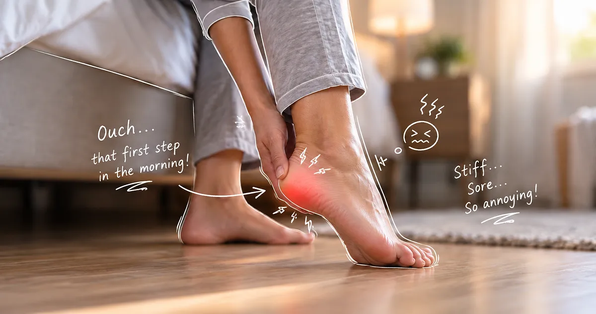
The pain is usually more central than the pain of plantar fasciopathy.
In a clinical study of 250 people with plantar heel pain, fat-pad atrophy accounted for approximately 14.8% of the diagnoses. Fat-pad pain was more likely to be bilateral, aggravated by standing and sometimes present at night. Plantar fasciopathy was more strongly associated with first-step morning pain and tenderness at the medial calcaneal tubercle (Yi et al., 2011).
These patterns are useful, but they are not absolute. A person can have both plantar fasciopathy and heel-pad dysfunction at the same time.
Heel fat pad syndrome versus plantar fasciopathy
These conditions are frequently confused because the plantar fascia and fat pad occupy neighbouring regions.
Plantar fasciopathy is more likely when:
Pain is concentrated around the medial plantar heel, particularly near the fascial attachment to the calcaneus. The first few steps after sleep or prolonged sitting are often especially painful.
Stretching the plantar fascia by lifting the toes may reproduce symptoms.
Heel fat pad dysfunction is more likely when:
Pain is centred directly beneath the calcaneus and feels like a deep bruise. Hard surfaces, barefoot walking and prolonged standing are prominent triggers.
Direct compression of the centre or margins of the heel pad may reproduce the pain.
Both may be present
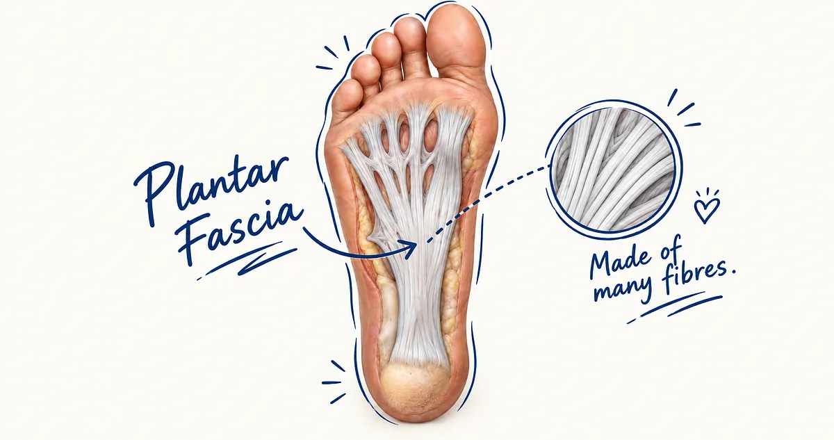
The fat pad lies superficial to the plantar fascia. A patient can therefore have fascial degeneration, heel-pad fibrosis, altered fat-pad compression and calcaneal pain simultaneously.
This is one reason that a diagnosis based solely on the word heel pain is inadequate.
How are heel fat pad problems detected?
Diagnosis begins with the history and physical examination.
A clinician should consider:
- the precise location of pain;
- whether pain is worse on first steps or after prolonged loading;
- sensitivity to hard surfaces;
- recent changes in exercise or footwear;
- direct injury;
- previous injections or surgery;
- night pain;
- neurological symptoms;
- and whether symptoms are unilateral or bilateral.
The foot should be examined both sitting and standing. The clinician may assess heel-pad thickness, displacement, tenderness, mobility and the ability of the tissue to remain centred beneath the calcaneus.
Ultrasound
Diagnostic ultrasound can show:
- unloaded thickness;
- thickness under controlled compression;
- superficial and deep layer behaviour;
- altered echogenicity;
- fibrosis;
- oedema;
- fluid crossing disrupted septa;
- abnormal vascularity;
- and dynamic subluxation.
A compressibility index may be calculated by comparing loaded and unloaded thickness. However, different studies use different methods, loads and formulae, making universal cut-off values difficult to establish.
Ultrasound is especially informative when the heel is examined dynamically rather than only in a relaxed, non-weight-bearing position.
MRI
MRI can provide a broader assessment of:
- fat-pad architecture;
- septal disruption;
- oedema and fibrosis;
- calcaneal stress injury;
- plantar fascia pathology;
- soft-tissue masses;
- and deeper inflammatory disease.
MRI may be appropriate when ultrasound is inconclusive or when bone, tumour, infection or extensive soft-tissue pathology needs to be excluded.
Why an abnormal scan does not necessarily mean disease
Imaging must be interpreted alongside the patient’s symptoms.
One ultrasound study found heel-pad echotexture abnormalities in 86% of the heels of asymptomatic endurance runners. Doppler blood flow was also visible in many painless heels (Hall et al., 2015).
This does not mean ultrasound is unreliable. It means that biological tissues develop adaptations and variations that may look unusual without causing pain.
A scan should not be treated as the diagnosis. The meaningful question is whether the imaging finding corresponds with:
- the painful location;
- the loading behaviour;
- the clinical examination;
- and the patient’s functional limitations.
How can you look after the heel fat pad?
There is no method that guarantees prevention of age-related change. Nevertheless, the mechanical environment surrounding the pad can be improved.
1. Avoid sudden increases in impact exposure
A fat pad accustomed to moderate walking may not immediately tolerate repeated jumping, sprinting or high-volume running.
Training load should increase gradually enough for the heel pad, skin, bone, fascia and muscles to adapt.
2. Do not confuse protection with complete rest
Short-term unloading may be appropriate after an acute contusion or severe flare. Prolonged avoidance of weight bearing is not automatically protective and may contribute to deconditioning of the heel tissues.
The aim is usually to find a tolerable level of loading rather than eliminate all loading indefinitely.
3. Limit barefoot walking on hard floors during painful periods
Barefoot walking is not universally harmful. However, when the pad is bruised, fibrotic, thinned or highly compressible, walking on tile, concrete or other rigid surfaces may reproduce symptoms because the biological pad must perform almost all the cushioning work.
Indoor footwear can make a substantial difference in these cases.
4. Reduce repeated direct heel trauma
Repeated jumping in thin, hard footwear, landing directly on the heel or continuing impact activity despite progressive bruise-like pain may exceed the pad’s recovery capacity.
5. Maintain general metabolic and vascular health
The heel fat pad depends on living cells, blood vessels and connective tissue. Diabetes, vascular disease and other systemic conditions can alter plantar soft-tissue properties.
General physical activity, appropriate nutrition and management of metabolic disease support tissue health throughout the body. This should not be reduced to an appearance-focused target or an assumption that every person with heel pain must lose weight.
Body mass is one mechanical variable among many.
6. Be cautious with repeated corticosteroid injections
Corticosteroid injections may be used selectively around the plantar heel, but repeated or poorly positioned injections have been associated with plantar fascia weakening and local fat-pad atrophy.
The potential benefit should be weighed against tissue-specific risks, particularly when the source of pain has not been clearly established.
What type of shoe protects the heel fat pad?
Direct clinical trials specifically investigating footwear for confirmed heel fat pad syndrome remain limited. Shoe advice must therefore combine available evidence with mechanical reasoning and individual assessment.
A suitable shoe commonly has several characteristics.
Adequate heel cushioning
The midsole should reduce the rate and magnitude of local heel compression. Cushioning should still rebound rather than remaining permanently flattened.
An old shoe may look intact externally while the heel foam has already lost much of its resilience.
A stable rearfoot platform
Extremely soft cushioning is not automatically superior.
If the heel sinks unevenly into an unstable midsole, the fat pad may experience additional shear and side-to-side displacement. The ideal shoe should combine cushioning with sufficient rearfoot stability.
A contoured heel seat
A gently cupped heel platform may help contain the pad beneath the calcaneus rather than allowing it to spread excessively beyond the heel margins.
Sufficient internal depth
Adding a heel cup or cushioning insert can raise the foot inside the shoe. There must be enough rearfoot depth to avoid heel slippage or pressure from the shoe collar.
Cushioning beneath the centre of the heel
A shoe that is soft only around its outer edge but firm directly beneath the calcaneus may not adequately protect the painful region.
Suitability for the activity and person
Walking, running, prolonged standing and court sports create different loading patterns. Body mass, foot posture, heel width, gait and the location of tissue damage also influence shoe choice.
The most appropriate shoe is therefore not necessarily the softest or most expensive one. It is the shoe that reduces symptoms while maintaining stable, comfortable movement.
Can heel cups, taping and orthoses help?
A heel cup can provide two potential benefits:
- it adds external cushioning; and
- it may help contain and centre the existing fat pad.
This is different from merely placing a flat piece of soft material beneath the heel. Containment may reduce excessive sideways expansion and improve the way pressure is distributed.
Taping can also temporarily reposition or contain the pad. In a clinical study involving patients with heel-pad atrophy, Low-Dye and modified Low-Dye taping reduced hindfoot peak pressure and pain, with the modified technique showing a greater immediate effect (Chae et al., 2018).
Taping is not a permanent replacement for healthy tissue. It can, however, serve as:
- a short-term pain-control measure;
- a way to test whether containment helps;
- or a bridge while footwear and activity are being modified.
An orthosis may help where foot mechanics repeatedly shift pressure onto a damaged part of the heel. It should not merely be prescribed because heel pain is present; its design should address the person’s specific loading pattern.
Can a damaged heel fat pad regrow?
The answer depends on what has been damaged.
A mild contusion, oedema or temporary disturbance of the tissue may improve as loading is modified and the tissue recovers. The heel pad remains vascular and biologically active.
Established atrophy, severe septal fragmentation or marked fibrosis is less likely to return completely to its original anatomy through exercise or footwear alone.
Autologous fat grafting—transferring a person’s own fat into the heel—is being studied for symptomatic fat-pad atrophy. A prospective randomised crossover trial reported improvements in pain and function following heel fat grafting, although the procedure remains specialised and questions remain regarding patient selection, long-term fat retention and restoration of normal chamber architecture (James et al., 2021).
Injecting loose fat does not automatically recreate the elaborate microchamber and macrochamber arrangement of a natural heel pad. An increase in volume and an improvement in symptoms do not necessarily mean that the original anatomy has been fully regenerated.
Conservative mechanical protection therefore remains the first approach for most patients.
What are the small fat lobules that appear when some people stand?
Some people notice multiple small, skin-coloured or yellowish bumps around the inner, outer or posterior margins of the heel when they stand.
These are commonly called piezogenic pedal papules.
The word piezogenic means produced by pressure.
When body weight compresses the heel, small portions of subcutaneous fat can protrude through weak points or gaps within the supporting connective tissue. The papules usually become less visible or disappear when weight is removed.
They are not tumours, and they are usually not a sign that dangerous fat is accumulating.
Are they normal?
Piezogenic papules are commonly found in healthy people, although reported prevalence varies enormously. The variation—from a few percent in some studies to the majority of examined participants in others—probably reflects differences in how carefully the heels were compressed and inspected.
In a study of 100 people, 80 had pedal papules. All were painless, multiple and bilateral. Biopsies demonstrated subcutaneous fat protruding into the lower dermis, together with fragmentation of dermal elastic fibres (Zaidi et al., 1995).
They have also been documented in healthy children (van Straaten et al., 1991).
Painless papules that appear only while standing and disappear when sitting are generally benign.
When can they become painful?
Painful piezogenic papules are much less common.
Pain may occur when protruding tissue places tension or compression on small vessels and nerves, or when the supporting septa have become damaged.
They may be more prominent in people exposed to frequent heel loading and in some connective-tissue disorders. Painful papules have been reported more frequently in Ehlers–Danlos syndrome, although the presence of painless papules alone does not establish a connective-tissue diagnosis (Kahana et al., 1987).
When should heel lumps be assessed?
Assessment is sensible when a lump:
- remains visible when the foot is unloaded;
- is firm rather than compressible;
- occurs only on one heel;
- is progressively enlarging;
- is red, warm, ulcerated or discoloured;
- produces significant pain;
- develops after an injury;
- or has an appearance inconsistent with ordinary pressure-induced papules.
Not every bump around the heel is a fat lobule. Cysts, bursae, vascular lesions, infection and soft-tissue tumours can also occur in this region.
Signs that your heel fat pad deserves examination
A professional assessment is appropriate when:
- heel pain persists despite reducing aggravating activities;
- the centre of the heel feels bruised for several weeks;
- pain is progressively worsening;
- there is night pain unrelated to pressure;
- a snapping or shifting sensation occurs beneath the heel;
- the pad appears displaced or markedly thinner;
- numbness, burning or tingling is present;
- there has been a significant fall or impact;
- the patient has diabetes, inflammatory disease or impaired circulation;
- or pain is preventing ordinary walking.
These features do not necessarily indicate serious disease. They indicate that plantar fasciitis should not be assumed without considering the fat pad, calcaneus, nerves and other soft tissues.
The heel fat pad deserves more respect
The contrast between visceral abdominal fat and heel fat demonstrates why it is misleading to regard all adipose tissue as the same.
Visceral adipose tissue is a large, biologically active depot capable of influencing glucose metabolism, vascular function and systemic inflammation when it becomes dysfunctional.
The heel fat pad is a small, highly reinforced and mechanically specialised depot. Its adipocytes are enclosed within a remarkable fibroelastic chamber system that:
- protects the calcaneus;
- distributes pressure;
- resists shear;
- adapts to different loading speeds;
- dissipates mechanical energy;
- and helps the foot interact safely with the ground.
The heel pad can age, bruise, stiffen, migrate, tear or become painful. It can also remain functional for a lifetime when its loading demands remain within its capacity.
Fat is therefore not inherently harmful.
Sometimes fat is a metabolic organ. Sometimes it is an energy reserve. Sometimes it fills spaces or supports delicate structures.
And beneath the heel, fat is an extraordinary piece of biological engineering that allows us to stand, walk and run without repeatedly striking the heel bone directly against the ground.
References
Balius, R., Bossy, M., Pedret, C., Porcar, C., Valle, X. and Corominas, H. (2021) ‘Heel fat pad syndrome beyond acute plantar fascitis’, The Foot, 48, 101829.
Buschmann, W.R., Jahss, M.H., Kummer, F., Desai, P., Gee, R.O. and Ricci, J.L. (1995) ‘Histology and histomorphometric analysis of the normal and atrophic heel fat pad’, Foot & Ankle International, 16(5), pp. 254–258.
Campanelli, V., Fantini, M., Faccioli, N., Cangemi, A., Pozzo, A. and Sbarbati, A. (2011) ‘Three-dimensional morphology of heel fat pad: an in vivo computed tomography study’, Journal of Anatomy, 219(5), pp. 622–631.
Chae, Y.H., Kim, J.S., Kang, M.H., Kim, H.M. and Jang, J.H. (2018) ‘Clinical and biomechanical effects of Low-Dye taping and figure-8 modification of Low-Dye taping in patients with heel pad atrophy’, Journal of Physical Therapy Science, 30, pp. 508–511.
De Clercq, D., Aerts, P. and Kunnen, M. (1994) ‘The mechanical characteristics of the human heel pad during foot strike in running: an in vivo cineradiographic study’, Journal of Biomechanics, 27(10), pp. 1213–1222.
Falsetti, P., Frediani, B., Acciai, C., Baldi, F., Filippou, G., Galeazzi, M. and Marcolongo, R. (2006) ‘Ultrasonography and magnetic resonance imaging of heel fat pad inflammatory-oedematous lesions in rheumatoid arthritis’, Scandinavian Journal of Rheumatology, 35(6), pp. 454–458.
Grigoriadis, G., Newell, N., Carpanen, D., Christou, A., Bull, A.M.J. and Masouros, S.D. (2017) ‘Material properties of the heel fat pad across strain rates’, Journal of the Mechanical Behavior of Biomedical Materials, 65, pp. 398–407.
Hall, M.M., Finnoff, J.T., Sayeed, Y.A. and Smith, J. (2015) ‘Sonographic evaluation of the plantar heel in asymptomatic endurance runners’, Journal of Ultrasound in Medicine, 34(10), pp. 1861–1871.
Jahss, M.H., Michelson, J.D., Desai, P., Kaye, R., Reich, S., Kummer, F., Buschmann, W. and Watkins, F. (1992) ‘Investigations into the fat pads of the sole of the foot: anatomy and histology’, Foot & Ankle, 13(5), pp. 233–242.
James, I.B., Gusenoff, B.R., Wang, S., DiBernardo, G., Minteer, D. and Gusenoff, J.A. (2021) ‘A step in the right direction: a prospective randomized, controlled crossover trial of autologous fat grafting for rejuvenation of the heel’, Aesthetic Surgery Journal, 41(7), pp. NP959–NP971.
Kahana, M., Feinstein, A. and Tabachnic, E. (1987) ‘Painful piezogenic pedal papules in patients with Ehlers–Danlos syndrome’, Journal of the American Academy of Dermatology, 17, pp. 205–209.
Maemichi, T., Tsutsui, T., Matsumoto, M., Iizuka, S., Torii, S. and Kumai, T. (2020) ‘The relationship of heel fat pad thickness with age and physiques in Japanese’, Clinical Biomechanics, 80, 105110.
Maemichi, T., Matsumoto, M., Okunuki, T. and Kumai, T. (2024a) ‘Changes in functional characteristics of heel fat pad with age’, Clinical Biomechanics, 118, 106294.
Maemichi, T. et al. (2024b) ‘Functional morphologic changes of the heel fat pad and plantar fascia in patients with heel pain during weightbearing and nonweightbearing’, Foot & Ankle Orthopaedics, 9.
Maemichi, T. et al. (2024c) ‘Effect of height and weight on heel fat pad movements during loading and unloading’, Foot & Ankle Orthopaedics, 9.
Maemichi, T., Matsumoto, M., Tsutsui, T. and Kumai, T. (2026) ‘Effect of long-term non-weight bearing on the morphology and function of the heel fat pad’, Journal of Foot and Ankle Research.
Neeland, I.J., Ayers, C.R., Rohatgi, A.K. et al. (2013) ‘Associations of visceral and abdominal subcutaneous adipose tissue with markers of cardiac and metabolic risk in obese adults’, Obesity, 21(9), pp. E439–E447.
Ozdemir, H., Söyüncü, Y., Özgörgen, M. and Dabak, K. (2004) ‘Effects of changes in heel fat pad thickness and elasticity on heel pain’, Journal of the American Podiatric Medical Association, 94(1), pp. 47–52.
Preis, S.R., Massaro, J.M., Robins, S.J. et al. (2010) ‘Abdominal subcutaneous and visceral adipose tissue and insulin resistance in the Framingham Heart Study’, Obesity, 18(11), pp. 2191–2198.
Sussman, W.I., Park, D.J., Rucci, P.M. and Chen, Y.H. (2021) ‘Subluxing fractured plantar fat pad: a case series and description of novel sonographic findings’, Skeletal Radiology, 50(6), pp. 1241–1247.
Svensson, H., Odén, B., Edén, S. and Lönn, M. (2014) ‘Adiponectin, chemerin, cytokines, and dipeptidyl peptidase 4 are released from human adipose tissue in a depot-dependent manner’, BMC Endocrine Disorders, 14, 7.
Van Straaten, E.A., van Langen, I.M. and Kuijpers, A.L.A. (1991) ‘Piezogenic papules of the feet in healthy children and their possible relation to connective tissue disorders’, Pediatric Dermatology.
Wearing, S.C., Smeathers, J.E., Yates, B., Urry, S.R. and Dubois, P. (2009) ‘Bulk compressive properties of the heel fat pad during walking: a pilot investigation in plantar heel pain’, Clinical Biomechanics, 24(4), pp. 397–402.
Yi, T.I., Lee, G.E., Seo, I.S., Huh, W.S., Yoon, T.H. and Kim, B.R. (2011) ‘Clinical characteristics of the causes of plantar heel pain’, Annals of Rehabilitation Medicine, 35(4), pp. 507–513.
Zaidi, Z., Jafri, N., Noori, B. and Thawerani, H. (1995) ‘Piezogenic papules: a study of 100 cases’, Journal of the Pakistan Medical Association, 45, pp. 93–94.
Frequently Asked Questions About the Heel Fat Pad
What is the heel fat pad?
The heel fat pad is a specialised fibro-adipose structure located between the plantar skin and the calcaneus, or heel bone. It is composed of fat cells enclosed within a reinforced network of collagenous and elastic partitions.
These partitions divide the tissue into smaller superficial microchambers and larger, more deformable deep macrochambers. This organised structure makes heel fat mechanically different from ordinary, relatively mobile subcutaneous fat elsewhere in the body (Jahss et al., 1992; Buschmann et al., 1995).
What does the heel fat pad do?
The heel fat pad helps distribute pressure, attenuate impact and protect the calcaneus during standing, walking and running.
It is a nonlinear viscoelastic tissue. This means it is relatively compliant during the early phase of compression but becomes progressively stiffer as it is loaded. Some of the mechanical energy applied to it is stored temporarily, while some is dissipated rather than being returned directly to the lower limb (Grigoriadis et al., 2017).
The heel pad is therefore more sophisticated than a simple cushion. Its architecture allows it to deform without normally collapsing completely beneath the heel.
What does heel fat pad pain feel like?
Heel fat pad pain is commonly felt as a deep, bruise-like pain directly beneath the centre of the heel. Some patients describe a sensation similar to walking on a stone or feeling as though the heel bone has insufficient cushioning.
Pain may be aggravated by prolonged standing, direct pressure beneath the heel and walking on hard surfaces. Fat-pad-related heel pain may also be bilateral and, in some patients, present at night (Yi et al., 2011; Chang et al., 2022).
These features can support the diagnosis, but none is completely specific. Calcaneal stress injury, nerve-related pain, inflammatory disease and plantar fasciopathy can produce overlapping symptoms.
How can I tell the difference between heel fat pad syndrome and plantar fasciitis?
Heel fat pad syndrome usually produces pain beneath the central part of the calcaneus. Direct compression of the middle of the heel often reproduces the symptoms, and prolonged standing may be particularly uncomfortable.
Plantar fasciopathy more commonly causes tenderness around the medial plantar heel, close to the plantar fascia’s attachment to the medial calcaneal tubercle. Pain during the first few steps after waking or after a period of sitting is more characteristic of plantar fasciopathy.
In a clinical study of 250 people with plantar heel pain, first-step morning pain and medial calcaneal tenderness were associated with plantar fasciopathy, whereas bilateral pain, night pain and pain aggravated by standing were more strongly associated with heel fat pad atrophy (Yi et al., 2011).
The conditions can coexist, so the distinction should not be based on one symptom alone.
Can the heel fat pad wear out?
The heel fat pad can deteriorate, but current evidence does not show that normal walking simply uses it up in the way that repeated driving wears down a tyre.
With age, injury or disease, the pad may become thinner, stiffer, more fibrotic, displaced or less capable of returning to its original shape after compression. Histological examinations of atrophied heel pads have demonstrated smaller fat cells and disruption or widening of the fibrous septa that normally contain the tissue (Buschmann et al., 1995).
Importantly, the scientific literature on heel fat pad syndrome remains limited. A 2022 scoping review identified only seven original studies meeting its criteria and found substantial gaps in the evidence concerning causes, diagnostic standards and conservative treatment (Chang et al., 2022).
It is therefore more accurate to say that heel pads can undergo structural and functional deterioration than to claim that everyone gradually “wears out” their heel fat.
Does the heel fat pad become thinner with age?
Heel fat pad thickness changes with age, but ageing does not always produce straightforward thinning.
In a study of 1,126 healthy participants aged from infancy to 96 years, heel fat pad thickness increased up to approximately 30–44 years of age and then gradually decreased in older groups. Thickness was also associated with body size and sex (Maemichi et al., 2020).
Other studies have found that older adults can have relatively thick heel pads that deform differently under load. Increased thickness may reflect changes in fat distribution, tissue composition or fibrosis rather than improved cushioning. Age can therefore alter both the structure and mechanical response of the microchamber and macrochamber layers (Ozdemir et al., 2004; Maemichi et al., 2024a).
A thicker heel pad is not necessarily a healthier heel pad.
How long should a healthy heel fat pad last?
There is no established lifespan or expiry date for the heel fat pad. It can remain functional throughout life, although its mechanical properties may change with ageing, injury, loading exposure and general health.
The available research is mostly cross-sectional, meaning that researchers compare people of different ages at one point in time. There are few long-term studies following the same healthy heel pads over several decades.
It is therefore not scientifically possible to state that a heel fat pad normally lasts a particular number of years. Its condition is better assessed through symptoms, clinical examination, tissue position, compressibility and load-dependent behaviour than through age alone (Maemichi et al., 2020; Maemichi et al., 2024a).
Does walking barefoot on hard floors wear down or permanently deform the heel fat pad?
Barefoot loading causes the heel pad to deform, but deformation during a step is not the same as permanent tissue damage.
In an in-vivo study of running, the heel pad underwent substantially greater compression during barefoot running than during shod running. Maximum percentage deformation averaged approximately 60.5% when barefoot and 35.5% when shoes were worn (De Clercq, Aerts and Kunnen, 1994).
However, this was a study of running, not ordinary household walking. It measured immediate deformation during loading; it did not demonstrate that barefoot walking on hard floors permanently thins, stretches or destroys a healthy heel pad.
At present, there is insufficient longitudinal evidence to conclude that normal barefoot walking on hard flooring inevitably wears away the heel fat pad. It is nevertheless plausible that prolonged or suddenly increased barefoot exposure could provoke symptoms when the pad is already bruised, fibrotic, thinned or mechanically dysfunctional.
Read next: Does Walking Barefoot on Hard Floors Damage or Wear Down the Heel Fat Pad?
That article will examine the difference between temporary deformation, tissue adaptation, repetitive overload and permanent structural change.
Can standing on hard floors damage the heel fat pad?
Standing on a hard floor can aggravate a painful heel pad, but it has not been proven that ordinary standing on hard flooring directly causes permanent fat-pad atrophy.
A rigid surface provides little external deformation, so cushioning must be supplied by the heel pad, footwear and the rest of the lower limb. Prolonged standing can therefore increase the duration of heel loading and may provoke pain when the tissue’s current capacity is exceeded.
However, the 2022 scoping review of heel fat pad syndrome found insufficient high-quality evidence to establish precise causal relationships between occupational standing, hard surfaces and heel fat pad degeneration (Chang et al., 2022).
Pain on hard floors should consequently be interpreted as a sign of reduced load tolerance rather than automatic proof that the pad has been permanently worn away.
Can heel fat pad atrophy heal or grow back?
There is currently no strong evidence that established heel fat pad atrophy spontaneously regenerates its original volume and chamber architecture.
Symptoms may improve when pressure is reduced and the pad is better protected, particularly when pain is related to a reversible contusion, inflammation or temporary overload rather than advanced structural atrophy. However, symptom improvement does not necessarily prove that lost fat or damaged septa have regrown.
Autologous fat grafting has been studied as a method of restoring volume. In a prospective randomised crossover trial, fat grafting improved patient-reported pain and foot function in people with heel fat pad atrophy (James et al., 2021).
Fat grafting remains a specialised intervention. Injected fat may improve cushioning, but it does not necessarily recreate the natural microchamber and macrochamber organisation of an undamaged heel pad.
What are the best shoes for heel fat pad pain?
No high-quality clinical trial has identified one particular shoe type, cushioning material or brand as the best treatment for heel fat pad syndrome.
From a biomechanical perspective, footwear may reduce how much the natural heel pad must compress. The barefoot-versus-shod running study by De Clercq, Aerts and Kunnen (1994) demonstrated considerably lower heel-pad deformation while participants were wearing shoes.
A suitable shoe would therefore generally be expected to provide cushioning directly beneath the heel while maintaining sufficient rearfoot stability. However, extremely soft footwear is not automatically better, particularly if it allows excessive heel movement or instability.
The evidence supporting specific footwear prescriptions remains sparse, and shoe recommendations should be presented as mechanically reasoned clinical management rather than as a treatment conclusively established by randomised trials (Chang et al., 2022).
Do heel cups or taping help heel fat pad syndrome?
Heel cups and taping may help by cushioning the heel and limiting excessive sideways displacement of the fat pad, but the evidence base is small.
A clinical study involving patients diagnosed with heel pad atrophy found that Low-Dye taping and a figure-of-eight modification reduced hindfoot peak pressure and immediate pain. The modified technique produced a greater reduction in pressure and symptoms than standard Low-Dye taping (Chae et al., 2018).
This provides preliminary support for externally containing or repositioning the heel tissues. It does not prove that taping repairs the fat pad, and the study assessed immediate effects rather than long-term tissue restoration.
Heel cups are frequently recommended on the basis of a similar containment principle, but robust clinical trials specifically evaluating heel cups for confirmed heel fat pad syndrome are lacking (Chang et al., 2022).
Can an ultrasound scan detect heel fat pad problems?
Ultrasound can help assess heel pad thickness, internal appearance, compressibility and behaviour during loading. It may also demonstrate oedema, fibrosis, septal disruption or abnormal movement.
Dynamic ultrasound is especially valuable because the superficial microchamber and deeper macrochamber layers do not behave identically. A 2024 study found that the macrochamber layer changed substantially during loading and unloading, while the microchamber layer remained comparatively stable. Patients with heel pain also showed greater deep-pad compression and less complete recovery after load removal (Maemichi et al., 2024b).
However, an abnormal ultrasound image does not necessarily identify the cause of pain. In a study of asymptomatic endurance runners, heel-pad echotexture abnormalities were found in 86% of examined heels, and Doppler flow was present in 88% (Hall et al., 2015).
Ultrasound findings must therefore be interpreted alongside the location of pain, loading history and clinical examination.
How thick should a normal heel fat pad be?
There is no universal heel fat pad thickness that is normal for every person.
In asymptomatic endurance runners, Hall et al. (2015) reported an average uncompressed thickness of approximately 13.7–13.8 mm. A much larger study showed that thickness also varies with age, height, body weight and sex (Maemichi et al., 2020).
Differences in ultrasound technique, probe pressure, anatomical measurement point and whether the foot is loaded can also change the result.
A single thickness measurement should therefore not be used to diagnose fat pad atrophy in isolation. Comparison with the opposite heel, assessment under controlled load and evaluation of the internal chamber structure may be more informative.
What are the small bumps that appear around the heel when standing?
Small bumps that appear around the heel during weight bearing are often piezogenic pedal papules.
They develop when pressure causes small portions of subcutaneous fat to protrude into or through weak points in the overlying connective tissue. They are commonly seen around the medial, lateral or posterior margin of the heel and usually become less prominent when the person sits or lifts the foot from the ground (Zaidi et al., 1995; van Straaten et al., 1991).
Piezogenic papules can occur in healthy adults and children. Their presence alone does not mean that the entire heel pad is failing or that the person has a connective-tissue disorder.
Are piezogenic heel papules dangerous?
Most piezogenic pedal papules are benign and painless.
In a study of 100 people, 80 demonstrated pedal papules, all of which were painless, multiple and bilateral. Histological examination showed subcutaneous fat protruding into the lower dermis with changes in the supporting elastic tissue (Zaidi et al., 1995).
They can occasionally become painful, possibly because the protruding tissue compresses or stretches small nerves and blood vessels. Painful papules have been reported in association with Ehlers–Danlos syndrome, where connective tissue is structurally more vulnerable (Kahana et al., 1987).
The finding of painless papules, however, is common and is not by itself evidence of Ehlers–Danlos syndrome.
When should bumps around the heel be examined?
Heel bumps should be assessed when they are painful, progressively enlarging, present on only one side or remain visible when the foot is completely unloaded.
Assessment is also advisable when a lesion is firm, discoloured, warm, ulcerated or appeared following a significant injury. Classic piezogenic papules are pressure dependent and normally become much less visible when weight is removed (van Straaten et al., 1991; Kahana et al., 1987).
A lump that does not behave in this way may have another explanation and should not automatically be assumed to represent harmless fat lobulation.
Can a corticosteroid injection damage the heel fat pad?
Local fat atrophy is a recognised potential complication of corticosteroid injection, but the risk appears to depend on the injection location, dose, frequency and technique.
Reviews of plantar heel pain treatment continue to identify plantar fascia rupture and fat pad atrophy as possible complications, particularly where injections are repeated or inadvertently placed into superficial fat (Latt et al., 2020).
However, the risk should not be overstated. In a long-term observational study of patients with plantar fasciopathy, mean heel pad thickness did not differ significantly between patients who had received an ultrasound-guided corticosteroid injection and those who had not. The authors found no evidence of heel fat pad atrophy attributable to the guided injections in their cohort (Hansen et al., 2018).
The balanced conclusion is that fat-pad atrophy is possible, particularly after inaccurate or repeated injections, but it is not an inevitable consequence of one appropriately positioned ultrasound-guided injection.
When should I see a podiatrist about possible heel fat pad pain?
An assessment is advisable when central heel pain persists, becomes progressively worse or limits ordinary walking and standing.
Earlier assessment is particularly appropriate when there is:
- pain following a fall or direct impact;
- significant swelling or bruising;
- pain at night that is not clearly related to pressure;
- numbness, burning or altered sensation;
- a snapping or shifting sensation beneath the heel;
- a visible change in the position of the heel pad;
- diabetes or reduced circulation;
- or failure to improve after sensible modification of aggravating activity.
Heel fat pad syndrome is only one possible cause of plantar heel pain. Plantar fasciopathy, calcaneal stress injury, inflammatory disease, nerve entrapment and other disorders may produce similar symptoms, making accurate differential diagnosis important (Yi et al., 2011; Chang et al., 2022).
References
Buschmann, W.R., Jahss, M.H., Kummer, F., Desai, P., Gee, R.O. and Ricci, J.L. (1995) ‘Histology and histomorphometric analysis of the normal and atrophic heel fat pad’, Foot & Ankle International, 16(5), pp. 254–258.
Chae, Y.H., Kim, J.S., Kang, Y., Kim, H.Y. and Yi, T.I. (2018) ‘Clinical and biomechanical effects of Low-Dye taping and figure-8 modification of Low-Dye taping in patients with heel pad atrophy’, Annals of Rehabilitation Medicine, 42(2), pp. 222–228.
Chang, A.H., Rasmussen, S.Z., Jensen, A.E. et al. (2022) ‘What do we actually know about a common cause of plantar heel pain? A scoping review of heel fat pad syndrome’, Journal of Foot and Ankle Research, 15, 60.
De Clercq, D., Aerts, P. and Kunnen, M. (1994) ‘The mechanical characteristics of the human heel pad during foot strike in running: an in vivo cineradiographic study’, Journal of Biomechanics, 27(10), pp. 1213–1222.
Grigoriadis, G., Newell, N., Carpanen, D., Christou, A., Bull, A.M.J. and Masouros, S.D. (2017) ‘Material properties of the heel fat pad across strain rates’, Journal of the Mechanical Behavior of Biomedical Materials, 65, pp. 398–407.
Hall, M.M., Finnoff, J.T., Sayeed, Y.A. and Smith, J. (2015) ‘Sonographic evaluation of the plantar heel in asymptomatic endurance runners’, Journal of Ultrasound in Medicine, 34(10), pp. 1861–1871.
Hansen, L., Krogh, T.P., Ellingsen, T., Bolvig, L. and Fredberg, U. (2018) ‘Long-term prognosis of plantar fasciitis: a 5- to 15-year follow-up study of 174 patients with ultrasound examination’, Orthopaedic Journal of Sports Medicine, 6(3), 2325967118757983.
Jahss, M.H., Michelson, J.D., Desai, P., Kaye, R., Reich, S., Kummer, F., Buschmann, W. and Watkins, F. (1992) ‘Investigations into the fat pads of the sole of the foot: anatomy and histology’, Foot & Ankle, 13(5), pp. 233–242.
James, I.B., Gusenoff, B.R., Wang, S., DiBernardo, G., Minteer, D. and Gusenoff, J.A. (2021) ‘A step in the right direction: a prospective randomized, controlled crossover trial of autologous fat grafting for rejuvenation of the heel’, Aesthetic Surgery Journal, 41(7), pp. NP959–NP971.
Kahana, M., Feinstein, A., Tabachnic, E., Schewach-Millet, M. and Engelberg, S. (1987) ‘Painful piezogenic pedal papules in patients with Ehlers–Danlos syndrome’, Journal of the American Academy of Dermatology, 17(2), pp. 205–209.
Latt, L.D., Jaffe, D.E., Tang, Y. and Taljanovic, M.S. (2020) ‘Evaluation and treatment of chronic plantar fasciitis’, Foot & Ankle Orthopaedics, 5(1).
Maemichi, T., Tsutsui, T., Matsumoto, M., Iizuka, S., Torii, S. and Kumai, T. (2020) ‘The relationship of heel fat pad thickness with age and physiques in Japanese’, Clinical Biomechanics, 80, 105110.
Maemichi, T., Matsumoto, M., Okunuki, T. and Kumai, T. (2024a) ‘Changes in functional characteristics of heel fat pad with age’, Clinical Biomechanics, 118, 106294.
Maemichi, T., Matsumoto, M., Tsutsui, T. et al. (2024b) ‘Functional morphologic changes of the heel fat pad and plantar fascia in patients with heel pain during weightbearing and nonweightbearing’, Foot & Ankle Orthopaedics, 9.
Ozdemir, H., Söyüncü, Y., Özgörgen, M. and Dabak, K. (2004) ‘Effects of changes in heel fat pad thickness and elasticity on heel pain’, Journal of the American Podiatric Medical Association, 94(1), pp. 47–52.
Van Straaten, E.A., Van Langen, I.M., Oorthuys, J.W.E. and Oosting, J. (1991) ‘Piezogenic papules of the feet in healthy children and their possible relation with connective tissue disorders’, Pediatric Dermatology, 8(4), pp. 277–279.
Yi, T.I., Lee, G.E., Seo, I.S., Huh, W.S., Yoon, T.H. and Kim, B.R. (2011) ‘Clinical characteristics of the causes of plantar heel pain’, Annals of Rehabilitation Medicine, 35(4), pp. 507–513.
Zaidi, Z., Jafri, N., Noori, B. and Thawerani, H. (1995) ‘Piezogenic papules—a study of 100 cases’, Journal of the Pakistan Medical Association, 45(4), pp. 93–94.