A Comprehensive Guide to Nail Disorders: What You Need to Know
9 June 2026
By Mark Reyneker, B.T. Pod (SA), MSc (SA)

Published: 9 June 2026 | Last reviewed: 9 June 2026
As we age, our bodies undergo numerous physiological changes, and our nails are no exception. Nail growth rates naturally decrease by approximately 0.5% per year starting at age 25, and nails often become dull, opaque, or pale over time (Abdullah and Abbas, 2011). While some nail alterations are a normal part of the aging process, many represent specific disorders that can cause severe pain, limit mobility, and significantly impact a person's quality of life (Abdullah and Abbas, 2011).
As we age, our bodies undergo numerous physiological changes, and our nails are no exception. Nail growth rates naturally decrease by approximately 0.5% per year starting at age 25, and nails often become dull, opaque, or pale over time (Abdullah and Abbas, 2011). While some nail alterations are a normal part of the aging process, many represent specific disorders that can cause severe pain, limit mobility, and significantly impact a person's quality of life (Abdullah and Abbas, 2011).
In this comprehensive guide, we will explore the most common nail disorders, their causes, and how they are typically managed.
Traumatic and Structural Nail Changes:
Onychogryphosis (Ram's Horn Nails)
Onychogryphosis is characterized by extreme thickening, hypertrophy, and a brown, opaque discoloration of the nail plate, most frequently affecting the great toenails (Albucker et al., 2024). The nail often curves upward and laterally, resembling a "ram's horn" or oyster shell, and is frequently caused by trauma, poor footwear, hallux valgus (bunions), or poor peripheral circulation (Albucker et al., 2024). Management typically involves mechanical debridement using electric files, chemical nail avulsion with urea, or surgical removal (Albucker et al., 2024).
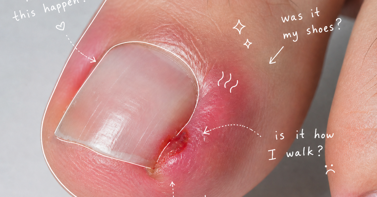
Onychocryptosis (Ingrown Toenails)
An ingrown toenail occurs when a spicule or edge of the nail plate penetrates the adjacent lateral nail fold, causing inflammation, pain, and secondary infection (Abdullah and Abbas, 2011). It is commonly caused by improper straight-line nail cutting, ill-fitting shoes, obesity, bony abnormalities, or trauma (Albucker et al., 2024). Mild cases are managed conservatively with warm soaks or taping, while severe cases may require surgical partial nail avulsion and matricectomy (Albucker et al., 2024).
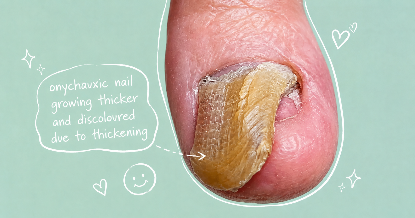
Onychauxis, also known as pachyonychia, presents as a localized thickening of the nail plate with a loss of translucency and subungual hyperkeratosis (Abdullah and Abbas, 2011). It is often triggered by advancing age, faulty biomechanics, or chronic pressure from overlapping toes (Abdullah and Abbas, 2011). Management includes regular filing, periodic debridement, or chemical nail avulsion (Albucker et al., 2024).
Onychophosis
This condition involves the hyperkeratosis (thickening of the skin) in the lateral or proximal nail folds, or in the subungual space between the nail fold and the nail plate (Albucker et al., 2024). It most often affects the first and fifth toes due to chronic shoe pressure and trauma (Albucker et al., 2024). Preventative care involves wearing comfortable shoes, while treatment focuses on debridement and applying keratolytics like urea or salicylic acid to soften the tissue (Albucker et al., 2024).
Onychoclavus (Subungual Corn)
An onychoclavus is a subungual heloma, or a "corn," that develops under the distal margin of the nail plate (Abdullah and Abbas, 2011). It manifests as a tender, dark area that can sometimes be mistaken for a subungual melanoma (Abdullah and Abbas, 2011). It is usually the result of chronic minor trauma or bony abnormalities like hammer toes (Albucker et al., 2024). Treatment consists of surgically removing the hyperkeratotic tissue and correcting any underlying bone issues (Abdullah and Abbas, 2011).
Book An Appointment
Book An Appointment
Pigment and Blood Abnormalities
Subungual Hematoma
A subungual hematoma is a collection of blood trapped underneath the nail, initially presenting as a painful, violaceous to black discoloration (Albucker et al., 2024). It is most commonly caused by acute trauma to the nail bed (Albucker et al., 2024). A key diagnostic feature is that the discolored spot will migrate distally (forward) as the nail grows, distinguishing it from melanoma (Abdullah and Abbas, 2011). Painful acute cases may require a doctor to drill a small hole in the nail to drain the blood and relieve pressure (Abdullah and Abbas, 2011).
Splinter Hemorrhages
These are thin, linear, dark brown or black lines beneath the nail plate (Albucker et al., 2024). When located in the middle or distal (end) third of the nail, they are typically the result of minor trauma (Abdullah and Abbas, 2011). However, if they appear in the proximal (base) area of the nail, they may be a warning sign of systemic diseases, such as bacterial endocarditis, connective tissue disorders, or nail psoriasis (Abdullah and Abbas, 2011).
Longitudinal Melanonychia
This is characterized by a brown to black band extending the length of the nail plate (Lee and Lipner, 2022). While it can be caused by benign melanocytic activation (due to ethnic variations, pregnancy, or medications) or benign nevi, it is also a hallmark sign of subungual melanoma, necessitating careful evaluation (Lee and Lipner, 2022).
Growth Disruptions
Beau's Lines
Beau's lines are transverse grooves that run across the nail plate caused by a temporary decrease in the mitotic activity of the nail matrix (Lee and Lipner, 2022). They occur due to a sudden shock to the system, such as a severe illness, systemic stress, physical trauma, or chemotherapy (Lee and Lipner, 2022). The distance of the groove from the cuticle can even help estimate the timeframe of the initial insult (Lee and Lipner, 2022).
Onychomadesis
Considered a more extreme presentation of Beau's lines, onychomadesis occurs when nail production halts completely, leading to the complete separation and shedding of the nail plate (Lee and Lipner, 2022). It is associated with severe illnesses (like Kawasaki disease or hand-foot-mouth disease), medications, and autoimmune conditions, and typically resolves on its own once the underlying factor is removed (Lee and Lipner, 2022).
Retronychia
Retronychia occurs when the nail plate's longitudinal growth is disrupted, causing the new nail to grow underneath the old one while remaining partially attached (Lee and Lipner, 2022). Instead of growing outward, the old nail is pushed backward (proximally) into the nail fold, causing significant inflammation, pain, and overlapping nail layers (Lee and Lipner, 2022). It is often triggered by repetitive trauma and is commonly treated by surgical nail avulsion (Lee and Lipner, 2022).
Infectious Conditions
Onychomycosis (Fungal Nail Infection)
Accounting for roughly 50% of all nail disorders, onychomycosis is an extremely common fungal infection, especially in older adults (Lee and Lipner, 2022). It presents with yellow discoloration, thickening, onycholysis (separation), crumbling, and a buildup of debris under the nail (Lee and Lipner, 2022). The most common pathogens are dermatophytes like Trichophyton rubrum (Abdullah and Abbas, 2011). Treatment involves topical antifungals or oral medications like terbinafine, though older adults may experience lower cure rates due to slower nail growth and poorer circulation (Albucker et al., 2024).
Paronychia (Acute, Chronic, and Chemotherapy-Associated)
Paronychia is an infection or inflammation of the nail folds (Lee and Lipner, 2022).
- Acute Paronychia: Usually caused by a bacterial infection (like Staphylococcus aureus) following a minor trauma, presenting as a highly painful, red, and swollen nail fold, often with an abscess (Lee and Lipner, 2022).
- Chronic Paronychia: A longer-lasting inflammation often linked to frequent exposure to water and environmental irritants (Lee and Lipner, 2022). Patients present with red, swollen folds and a loss of the cuticle (Lee and Lipner, 2022).
- Chemotherapy-Associated Paronychia (CAP): Induced by chemotherapeutic agents like EGFR inhibitors and taxanes, this causes severe erythema and pain, requiring specific dermatological management (Lee and Lipner, 2022).
Periungual and Subungual Warts
Caused by the Human Papillomavirus (HPV), warts can form around or underneath the nail unit, particularly in individuals with a weakened immune system (Albucker et al., 2024). Because the nail plate protects the virus, they are notoriously challenging to treat and may require destructive therapies like cryosurgery, lasers, or intralesional injections (Albucker et al., 2024).
Inflammatory and Structural Disorders
Brittle Nail Syndrome (BNS)
Affecting up to 20% of the population, BNS is particularly common in older women and causes increased fragility of the nails (Lee and Lipner, 2022). It presents in two main forms: onychoschizia (transverse lamellar splitting and peeling at the tips) and onychorrhexis (longitudinal ridges and splitting) (Abdullah and Abbas, 2011). It is exacerbated by frequent handwashing, harsh cosmetics, and repetitive trauma (Abdullah and Abbas, 2011). Treatment involves intensive hydration with emollients rich in phospholipids, and sometimes oral biotin or silicon supplements (Abdullah and Abbas, 2011).
Nail Psoriasis
Psoriasis isn't just a skin condition; it can severely affect the nails, causing significant pain and impacting daily functions (Lee and Lipner, 2022). Signs of nail matrix psoriasis include deep pits, crumbling, and white spots (leukonychia), while nail bed psoriasis presents with "oil drop" or "salmon" patches, splinter hemorrhages, and onycholysis (Lee and Lipner, 2022). Treatment may involve topical steroids, intralesional steroid injections, or systemic biologic medications (Lee and Lipner, 2022).
Neoplastic (Growth) Changes and Cysts
Nail Unit Melanoma
Though rare, melanoma can develop under the nail, frequently peaking in patients aged 50 to 70 (Lee and Lipner, 2022). It classically presents as longitudinal melanonychia—a dark brown or black band running the length of the nail (Lee and Lipner, 2022). A highly concerning indicator is Hutchinson’s sign, where the pigment extends from the nail bed into the surrounding tissue of the nail folds (Abdullah and Abbas, 2011). Early diagnosis is critical, and treatment typically involves en bloc excision or amputation of the affected digit (Lee and Lipner, 2022).
Bowen’s Disease
Bowen's disease is a form of squamous cell carcinoma in situ that occasionally affects the nail unit, most commonly in men around age 70 (Albucker et al., 2024). Often associated with HPV infections, trauma, or radiation, it appears as an ulcerated, hyperkeratotic lesion with scaling and crusting (Abdullah and Abbas, 2011). The gold standard treatment is Mohs micrographic surgery to preserve digital function while removing the cancer (Abdullah and Abbas, 2011).
Digital Myxoid Cysts
Myxoid cysts are benign, translucent, dome-shaped nodules that form near the joints of the fingers and toes (Albucker et al., 2024). Because they are often linked to osteoarthritis, they are very common in older adults (Albucker et al., 2024). When located near the proximal nail fold, the cyst can compress the nail matrix, resulting in a distinct longitudinal groove running down the nail plate (Albucker et al., 2024). Asymptomatic cysts are usually observed, but symptomatic ones can be drained or surgically excised (Albucker et al., 2024).
Book An Appointment
Disclaimer: The word "treatment" in this article refers to the care and management of a patient’s health to prevent, cure, or improve a condition. Treatment results vary and do not necessarily indicate a cure. This article is for informational and educational purposes only and does not constitute medical advice.
About the author
Mark Reyneker is a podiatrist and human gait specialist with 8 years of training and over 25 years of clinical experience. He is the Founder and Clinical Director of Family Podiatry Centre and has a Bachelors degree in Podiatric Medicine and a Master’s degree in paleoanthropology, with research focused on human foot function and metatarsal loading.
References
1. Abdullah, L. and Abbas, O. (2011) 'Common nail changes and disorders in older people: Diagnosis and management', Canadian Family Physician, 57(2), pp. 173–181.
2. Albucker, S.J., Conway, J. and Lipner, S.R. (2024) 'Nails in older adults', Annals of Medicine, 56(1), p. 2336989.Lee, D.K. and Lipner, S.R. (2022) 'Optimal diagnosis and management of common nail disorders', Annals of Medicine, 54(1), pp. 694–712.
3. Lee, D.K. and Lipner, S.R. (2022) 'Optimal diagnosis and management of common nail disorders', Annals of Medicine, 54(1), pp. 694–712.