Overlapping Second Toe: The Hidden Link With Plantar Plate Injury, Forefoot Overload, Capsule Tears and Osteoarthritis
29 June 2026
By Mark Reyneker, B.T. Pod (SA), MSc (SA)
Published: 29 June 2026 | Last reviewed: 13 July 2026
An overlapping second toe is often treated as a cosmetic toe deformity, but in adults it is frequently a sign of deeper mechanical failure inside the forefoot. The toe does not usually cross over by accident. In many cases, it is the visible result of progressive instability at the second metatarsophalangeal joint, commonly called the second MTP joint.
An overlapping second toe is often treated as a cosmetic toe deformity, but in adults it is frequently a sign of deeper mechanical failure inside the forefoot. The toe does not usually cross over by accident. In many cases, it is the visible result of progressive instability at the second metatarsophalangeal joint, commonly called the second MTP joint.
This condition is closely linked with plantar plate injury, forefoot overload, capsulitis, collateral ligament damage, hammertoe formation, joint subluxation and, in long-standing cases, osteoarthritis. In the medical literature, similar problems may be described as crossover second toe, lesser metatarsophalangeal joint instability, predislocation syndrome, plantar plate insufficiency, plantar plate tear, or second MTP joint instability (Coughlin, 1987; Yu et al., 2002; Doty and Coughlin, 2014; Maas et al., 2016).
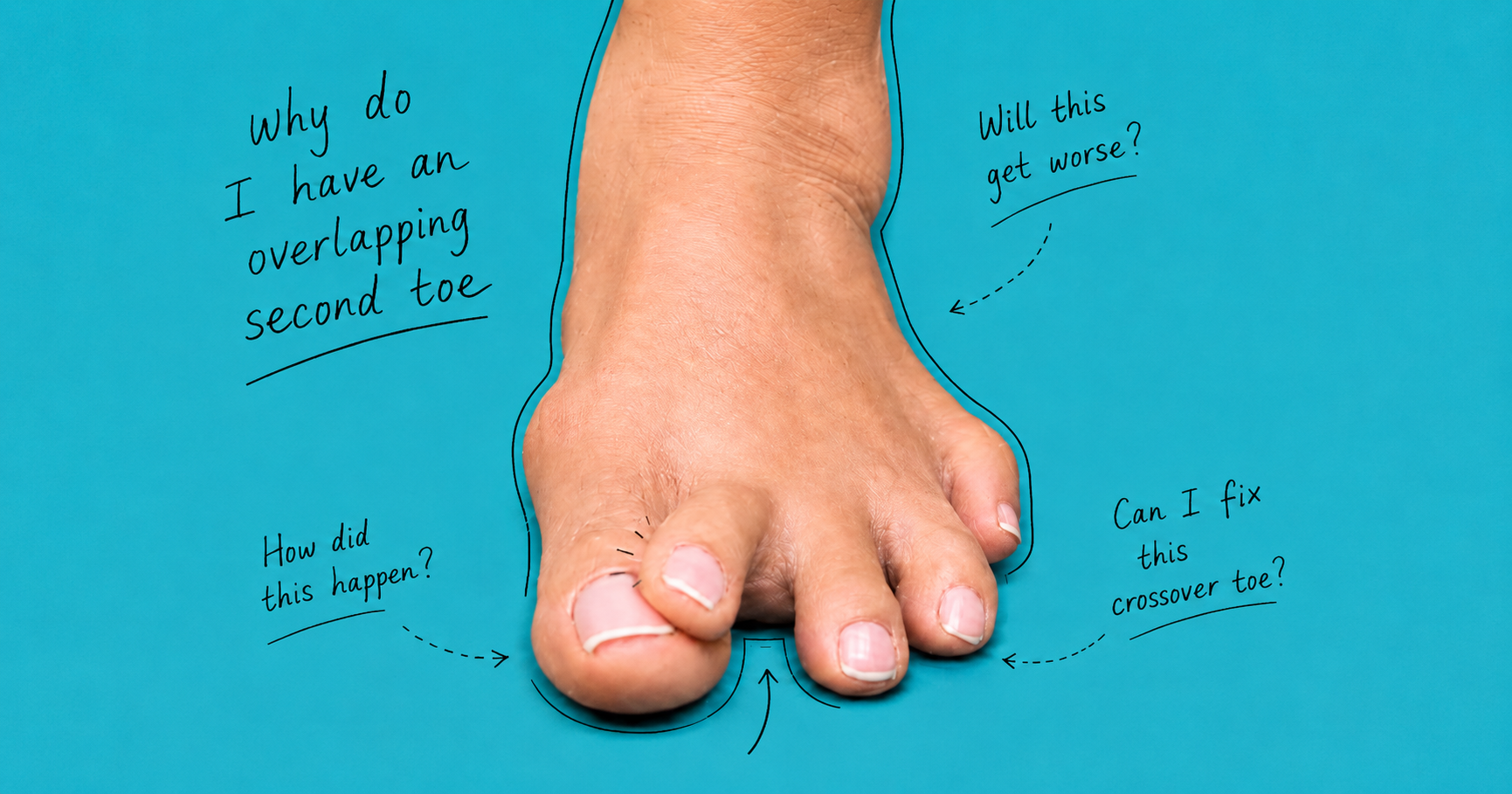
The photograph shown here demonstrates the type of severe second-toe overlap that clinicians often associate with advanced lesser MTP joint instability. A diagnosis, however, should not be made from appearance alone. A proper assessment usually includes clinical testing, weight-bearing X-rays and, where needed, ultrasound or MRI.
What is an overlapping second toe?
An overlapping second toe occurs when the second toe drifts out of its normal alignment and begins to sit over, across or against the big toe. In adults, this is often not simply a skin, nail or shoe-pressure problem. It commonly reflects a deeper loss of joint stability at the base of the toe.
The second toe is attached to the foot at the second metatarsophalangeal joint. This joint must remain stable while still allowing the toe to bend during walking. During push-off, the forefoot accepts high loads, and the lesser MTP joints must resist excessive upward bending, sideways drifting and dorsal displacement of the toes. When the stabilising structures fail, the toe may begin to lift, drift, hammer, cross over or dislocate.
Why the plantar plate is central to this condition
The plantar plate is a strong fibrocartilaginous structure located on the underside of the lesser metatarsophalangeal joints. It acts like a stabilising platform beneath the base of the toe. It helps resist excessive dorsiflexion of the toe and helps prevent the proximal phalanx from sliding upwards on the metatarsal head during walking (Deland et al., 1995; Johnston et al., 1994; Gregg et al., 2007).
Although people often talk about a “plantar plate tear” as though it is one isolated injury, the problem is usually more complex. The plantar plate blends functionally with the joint capsule, collateral ligaments, flexor tendon sheath and deep transverse metatarsal ligament. Therefore, when the plantar plate weakens or tears, the whole capsuloligamentous stabilising system of the joint may be compromised (Maas et al., 2016).
This is why the visible overlapping toe is often only the final external sign of a much deeper structural problem.
Why the second toe is commonly affected
The second MTP joint is especially vulnerable because it is often a major transfer-loading point in the forefoot. When the first ray and big toe joint do not accept load efficiently, pressure can shift towards the second metatarsal head. This may occur with hallux valgus, first-ray insufficiency, reduced big toe function, a relatively long second metatarsal, altered metatarsal parabola, high-heeled shoes, tight footwear or repetitive forefoot loading (Fleischer et al., 2017; Mann et al., 2021).
The second metatarsal is also relatively fixed compared with some of the other rays. This means that repeated overload beneath the second metatarsal head can concentrate stress around the second MTP joint. Over time, this may irritate the joint capsule, stretch the plantar plate, damage the collateral ligaments and allow the toe to drift.
In simple terms, an overlapping second toe often develops because the joint beneath the toe is no longer being held in a stable, centred position.
The link between forefoot overload and plantar plate injury
Forefoot overload is one of the most important mechanical drivers of lesser MTP joint instability. When excessive pressure is repeatedly placed under the second metatarsal head, the soft tissues beneath the joint are exposed to repetitive strain. At first, this may cause pain and inflammation. Later, it may lead to attenuation or tearing of the plantar plate.
Patients may describe pain under the ball of the foot, usually beneath the second toe joint. Some describe it as walking on a pebble, a bruised feeling, a burning discomfort, or a sense that the toe is unstable. These symptoms may be worse in flexible shoes, narrow shoes, high heels, during long walks, or during activities that increase forefoot loading.
This stage is important because the toe may still look relatively normal. In early plantar plate injury, pain and swelling may appear before the toe visibly crosses over. By the time the second toe is obviously overlapping, the condition is often more advanced.
The degenerative cascade: how an overlapping second toe develops
An overlapping or crossover second toe usually develops gradually. It is rarely an isolated cosmetic deformity. More often, it represents a progressive failure of the soft-tissue stabilisers of the second metatarsophalangeal joint, especially the plantar plate, collateral ligaments and joint capsule.
1. Early forefoot overload
The process often begins with excessive pressure beneath the second metatarsal head. This may happen when the first ray is not accepting load efficiently, when there is hallux valgus, when the second metatarsal is relatively long, or when footwear repeatedly increases forefoot pressure.
At this stage, the patient may feel pain under the ball of the foot, often described as walking on a pebble, bruising, burning, or discomfort directly beneath the second toe joint. The toe may still look reasonably straight, but the joint is already becoming irritated.
2. Synovitis and capsulitis
With continued overload, the second metatarsophalangeal joint may become inflamed. The synovial lining and capsule react to repetitive stress. This is often called capsulitis or synovitis.
Clinically, the joint may feel swollen, tender and sensitive to pressure. The patient may notice that symptoms are worse after prolonged walking, standing, running, wearing tight shoes, or using shoes with a higher heel-to-toe drop. At this stage, the deformity may still be subtle, but the joint is beginning to lose its normal mechanical environment.
3. Plantar plate attenuation
If the overload continues, the plantar plate may begin to stretch and weaken. This is known as attenuation. The plantar plate is no longer able to hold the base of the toe firmly against the metatarsal head during walking.
This is the stage where toe purchase may start to reduce. The toe may not press into the ground as strongly as the other toes. The patient may notice that the second toe is beginning to lift, drift, or feel unstable. A drawer test may become mildly positive.
4. Partial plantar plate tear
As degeneration progresses, the weakened plantar plate may develop a partial tear. This is often the transition from a painful but stable joint to a painful and mechanically unstable joint.
The toe may begin to separate from the neighbouring toes. There may be visible splaying, early hammering, or mild deviation of the second toe towards the big toe. Pain is usually more localised beneath the second metatarsophalangeal joint. Some patients describe a feeling that the toe is “not sitting properly”.
5. Full plantar plate rupture and capsular failure
In more advanced cases, the plantar plate may rupture more completely. The joint capsule and collateral ligaments may also fail. At this point, the second toe can no longer remain centred on the metatarsal head.
The toe may drift upwards, sideways, or across the big toe. This is when the classic crossover or overlapping second toe becomes more obvious. The drawer test is usually more clearly positive, and the toe often loses meaningful ground purchase.
6. MTP joint subluxation or dislocation
Once the plantar plate and capsule can no longer stabilise the joint, the base of the proximal phalanx may begin to sublux or dislocate on the metatarsal head. This means the joint surfaces are no longer aligned properly.
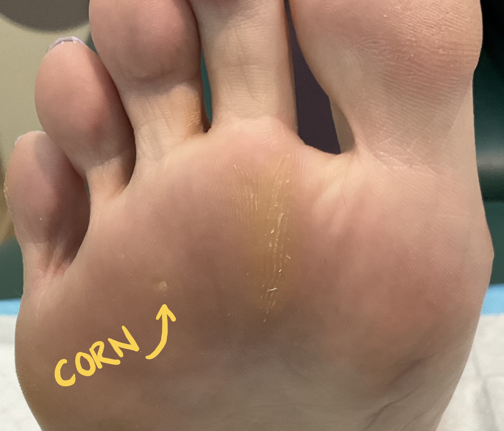
The toe may sit visibly out of position. Walking can become uncomfortable because load is no longer being transferred evenly through the forefoot. Corns, callus, shoe irritation and transfer metatarsalgia may become more prominent.
7. Degenerative osteoarthritis
In long-standing cases, the second metatarsophalangeal joint may develop degenerative change. Chronic malalignment means the cartilage surfaces are loaded unevenly. Over time, this can contribute to stiffness, joint-space narrowing, osteophyte formation and osteoarthritis.
At this stage, treatment becomes more complex. The problem is no longer only a soft-tissue injury. It is now a combined problem of plantar plate failure, capsular insufficiency, toe deformity, altered forefoot loading and joint degeneration.
Capsule tears and collateral ligament failure
The plantar plate does not fail alone. In advanced overlapping second toe deformity, the joint capsule and collateral ligaments are often involved. The capsule surrounds the joint, while the collateral ligaments help resist sideways movement. When these structures stretch or tear, the second toe can drift medially, dorsally or obliquely across the big toe.
This explains why some patients develop a progressive widening between the second and third toes before the second toe crosses over. Others develop a hammering pattern first, followed by sideways drift. The final appearance depends on which soft tissues fail first, how the metatarsal heads are loaded, and whether there is associated hallux valgus or first-ray insufficiency.
Crossover toe is therefore best understood as a three-dimensional deformity. It is not simply a toe moving sideways. It often involves dorsal subluxation, medial deviation, plantar plate failure, capsular insufficiency, extensor tendon imbalance and sometimes hammertoe contracture (Coughlin, 1987; Coughlin, 1993; Doty and Coughlin, 2014).
The role of osteoarthritis
Osteoarthritis can both contribute to and result from this process. In some patients, degenerative change within the second MTP joint may reduce joint congruity and increase capsular irritation. In others, the osteoarthritis develops later because the joint has been malaligned for a long time.
A chronically subluxed second MTP joint does not load normally. Instead of smooth, centred joint contact, the cartilage surfaces may be compressed unevenly. This can lead to joint-space narrowing, osteophytes, stiffness and persistent pain. Once osteoarthritis is established, treatment is less predictable because the condition is no longer just a soft-tissue instability. It has become a combined problem of deformity, overload and joint degeneration (Mann et al., 2022).
This is one reason early recognition matters. The earlier the plantar plate and capsule problem is identified, the more realistic it is to offload and stabilise the joint before fixed deformity and arthritis develop.
Clinical signs that suggest plantar plate injury
The most important clinical signs are functional rather than cosmetic.
Pain beneath the second metatarsal head is a common early symptom. Swelling around the second MTP joint may also be present. Patients often report gradual onset of pain rather than a single traumatic incident, although acute worsening can occur after a particular walk, run or change in footwear (Klein et al., 2013a).
One of the most important clinical tests is the drawer test, also called the vertical Lachman test. During this test, the clinician stabilises the metatarsal and checks whether the toe can be translated upwards excessively. Increased dorsal movement, especially without a firm endpoint, suggests plantar plate and capsular insufficiency.
Another useful observation is toe purchase. A healthy toe should be able to press down into the ground during standing and walking. When the plantar plate and flexor stabilising mechanism are compromised, the toe may lose its ability to grip the ground. Loss of toe purchase is often a sign that the stabilising system is no longer functioning properly.
Visible crossover deformity is usually a late sign. It is strongly suggestive of advanced instability when present, but its absence does not rule out a plantar plate injury. Many patients with early plantar plate pathology have pain and swelling before the toe visibly overlaps.
Imaging: X-ray, ultrasound and MRI
Weight-bearing X-rays are usually the starting point. They do not show the plantar plate directly, but they can show important indirect information. This includes metatarsal length pattern, hallux valgus, second MTP subluxation, osteoarthritis, Freiberg-type change, lesser toe deformity and the overall structure of the forefoot.
Ultrasound can be useful because it allows dynamic assessment. It may show thickening, thinning, discontinuity or abnormal motion of the plantar plate. It can also be used while stressing the joint. The limitation is that ultrasound is operator-dependent, and accuracy can vary depending on the skill and experience of the examiner (Duan et al., 2017; McCarthy and Thompson, 2021).
MRI is valuable because it provides a broader anatomical view. It can show plantar plate degeneration, partial tears, full-thickness tears, joint effusion, synovitis, bone marrow oedema, interspace lesions and osteoarthritic changes. MRI may also help distinguish plantar plate pathology from Morton’s neuroma, stress injury, inflammatory arthritis or other causes of forefoot pain (Ashman et al., 2001; Umans et al., 2014; Yamada et al., 2017).
A 2022 systematic review and meta-analysis found that both MRI and dynamic ultrasound can be useful for detecting lesser metatarsal plantar plate injuries, although each modality has strengths and limitations (Albright et al., 2022).
Why treatment must address the cause, not just the toe
Treatment should not focus only on making the toe look straighter. If the underlying overload remains, the joint may continue to deteriorate.
In early or flexible cases, conservative management usually focuses on reducing pressure beneath the painful metatarsal head and improving joint stability. This may include footwear changes, a wider toe box, stiff or rocker-type soles, reduction of high-heel use, metatarsal padding, plantar plate taping, toe splinting, orthoses and activity modification.
A metatarsal dome or pad is usually positioned just behind the painful metatarsal head rather than directly under it. The aim is to reduce peak pressure beneath the overloaded joint. Orthoses may be used to redistribute load, improve first-ray function where possible, and reduce excessive transfer pressure into the second metatarsal region.
Taping may help hold the toe in a more plantarflexed and aligned position during walking. This can be particularly useful in earlier stages, when the toe is still reducible and the joint has not become rigid.
In advanced cases, conservative treatment may reduce pain and improve shoe comfort but may not reverse the deformity. Once there is fixed crossover, severe subluxation, dislocation or osteoarthritis, the structural deformity may be too advanced for taping and orthoses alone.
Surgical considerations
Surgical treatment may be considered when pain, deformity and instability remain significant despite conservative care. Surgical options vary depending on the severity of the plantar plate tear, the degree of subluxation, the presence of hammertoe deformity, metatarsal length, hallux valgus, soft-tissue balance and joint degeneration.
Procedures discussed in the literature include direct plantar plate repair, Weil metatarsal osteotomy, capsular balancing, collateral ligament repair or reefing, hammertoe correction, flexor tendon transfer and correction of associated hallux valgus where it contributes to second-ray overload (Nery et al., 2012; Doty and Coughlin, 2014; Akoh and Phisitkul, 2018).
In arthritic or rigid cases, the decision-making becomes more complex. A joint-preserving procedure may be less predictable if the cartilage is already significantly damaged. This is why weight-bearing radiographs and, in selected cases, MRI can be useful before deciding on treatment direction.
Why early assessment matters
An overlapping second toe is often the late presentation of a problem that began much earlier. The early warning signs may be subtle: pain under the second metatarsal head, swelling around the second toe joint, reduced toe purchase, a feeling of instability, or mild separation between the second and third toes.
The earlier the condition is identified, the more opportunity there is to offload the joint, protect the plantar plate and reduce progression. Waiting until the toe is fully crossed over may limit conservative options and make treatment more complex.
Key message
An overlapping second toe should not be dismissed as a simple toe deformity. In adults, it is often linked to plantar plate injury, forefoot overload, capsular degeneration, collateral ligament failure and lesser metatarsophalangeal joint instability.
By the time the second toe visibly overlaps the big toe, the underlying joint may already have gone through a long process of inflammation, soft-tissue stretching, plantar plate tearing, subluxation and degenerative change. A careful assessment should therefore look beyond the toe itself and examine the whole forefoot loading pattern.
The goal is not only to straighten the toe. The real goal is to understand why the toe became unstable in the first place.
Book An Appointment
Disclaimer: The word "treatment" in this article refers to the care and management of a patient’s health to prevent, cure, or improve a condition. Treatment results vary and do not necessarily indicate a cure. This article is for informational and educational purposes only and does not constitute medical advice.
About the author
Mark Reyneker is a podiatrist and human gait specialist with 8 years of training and over 25 years of clinical experience. He is the Founder and Clinical Director of Family Podiatry Centre and has a Bachelors degree in Podiatric Medicine and a Master’s degree in paleoanthropology, with research focused on human foot function and metatarsal loading.
References
Recommended posts