Stiff Big Toe Joint: A Comprehensive Guide to Hallux Rigidus



11 July 2026
By Mark Reyneker, BTech (Podiatry), MSc (Palaeontology), Registered Podiatrist, Foot & Gait Specialist | Founder & Clinical Director, Family Podiatry Centre
When the big toe becomes stiff, painful and difficult to bend
What is hallux rigidus?
- pain at the base of the big toe;
- stiffness during walking;
- swelling around the joint;
- reduced ability to bend the toe upwards;
- pain during push-off;
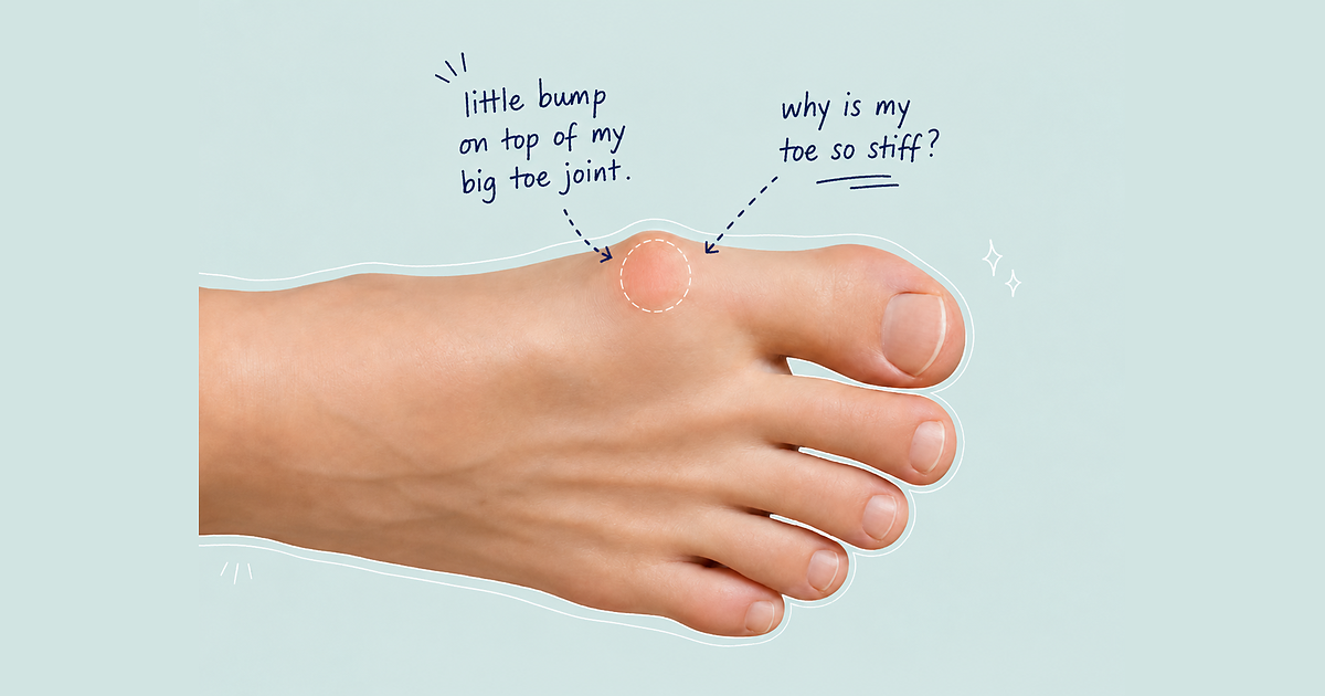
- a bony bump on top of the joint;
- difficulty wearing shoes;
- pain in flexible shoes;
- altered walking pattern; and
- progressive loss of joint motion.
Hallux rigidus is one of the most common arthritic problems of the foot and can significantly affect walking, exercise and quality of life (Bergin et al., 2012; Roddy et al., 2015).
Book An Appointment
Hallux rigidus, hallux limitus and functional hallux limitus
Hallux limitus
This is clinically important because a patient may say, “My toe bends when I pull it up with my hand, so why does it hurt when I walk?” The answer is that the joint may behave very differently under body weight.
Why the big toe joint is so important
The big toe joint is small, but it has a major role in walking and propulsion.
During walking, the heel contacts the ground, the foot accepts body weight, the body moves forwards, and the heel lifts. As the heel rises, the big toe must bend upwards. This movement is called dorsiflexion.
For efficient walking, the big toe joint needs:
- adequate dorsiflexion;
- smooth cartilage;
- good sesamoid movement;
- stable first-ray mechanics;
- appropriate plantar fascia tension;
- adequate ankle mobility;
- suitable footwear; and
- coordinated muscle control.
If the first metatarsal remains too elevated, if the joint is blocked by bone spurs, or if the foot mechanics prevent normal gliding, the big toe joint may jam during push-off. Repeated jamming is one of the central podiatric concepts in hallux rigidus.
What happens inside the joint?
- thinning of articular cartilage;
- roughening of the cartilage surface;
- inflammation of the synovial lining;
- thickening of the joint capsule;
- subchondral bone stress;
- subchondral sclerosis;
- cyst formation;
- flattening of the first metatarsal head;
- dorsal bone spur formation;
- reduced sesamoid movement;
- pain at end-range dorsiflexion; and
- progressive loss of movement.
The pathogenesis of hallux rigidus: how the condition develops
Hallux rigidus often develops through a chain reaction.
The cartilage may be injured by trauma, repetitive compression or abnormal loading. Once the cartilage surface becomes less smooth, the joint becomes less able to distribute pressure evenly.
During push-off, the base of the big toe may contact the top of the first metatarsal head too early. This creates dorsal joint compression and pain at the top of the joint.
Step 4: bone spurs form
Step 6: walking pattern changes
This self-reinforcing cycle explains why hallux rigidus may gradually progress from mild stiffness to significant arthritis.
What causes hallux rigidus?
- first metatarsal head shape;
- joint congruity;
- cartilage resilience;
- first-ray alignment;
- sesamoid position;
- hallux length;
- ligament stiffness; and
- the way the foot loads during gait.
- severe stubbing of the big toe;
- turf-toe injury;
- hyperextension injury;
- direct impact;
- fracture involving the joint;
- cartilage injury;
- sesamoid injury; or
- repetitive sports-related jamming.
3. Repetitive compression during push-off
Every step loads the big toe joint. Walking, running, climbing, lunging and squatting all require the joint to tolerate compression and dorsiflexion.
A joint may be vulnerable because of:
- reduced first-ray plantarflexion;
- metatarsus primus elevatus;
- previous cartilage injury;
- limited ankle dorsiflexion;
- poor footwear;
- joint incongruity; or
- functional hallux limitus.
Metatarsus primus elevatus means the first metatarsal sits higher than expected relative to the lesser metatarsals.
However, the evidence is debated.
This is one reason why not every patient with hallux rigidus should simply stretch the big toe harder. Some patients need better first-ray loading, improved shoe structure or orthotic control.
6. Soft-tissue restriction
Bone is not the only factor. Soft tissues may also restrict big toe motion.
- plantar fascia tension;
- capsular tightness;
- flexor hallucis longus restriction;
- sesamoid restriction;
- intrinsic muscle dysfunction;
- calf tightness;
- limited ankle dorsiflexion; and
- protective muscle guarding.
7. Abnormal first metatarsal head shape
- reduce smooth gliding;
- concentrate pressure;
- increase dorsal impingement;
- reduce tolerance to trauma; and
- encourage osteophyte formation.
Shoes that commonly aggravate the condition include:
- narrow shoes;
- shallow toe-box shoes;
- high heels;
- very flexible shoes;
- minimalist shoes;
- thin-soled shoes;
- shoes that bend directly under the painful joint;
- shoes that are too short; and
- shoes that rub the dorsal bone spur.
Not every stiff big toe is ordinary osteoarthritis.
- gout;
- rheumatoid arthritis;
- psoriatic arthritis;
- infection;
- osteochondral injury;
- sesamoiditis;
- stress fracture; and
- previous surgical complications.
Symptoms of hallux rigidus
- pain at the base of the big toe;
- stiffness during walking;
- pain during push-off;
- swelling around the joint;
- a hard bump on top of the joint;
- difficulty wearing shoes;
- pain in flexible footwear;
- discomfort walking uphill;
- difficulty with lunges or squats;
- pain after running or long walks;
- grinding or clicking;
- reduced stride length;
- walking on the outside of the foot;
- callus beneath the lesser metatarsals; and
- pain under the ball of the foot.
Why the top of the big toe joint becomes painful
The top of the joint is commonly painful because this is where dorsal impingement occurs.
- cartilage compression;
- synovial irritation;
- capsular inflammation;
- bone spur formation;
- shoe rubbing; and
- pain when the toe is pushed upwards.
Book An Appointment
How hallux rigidus changes walking
Canseco et al. (2008) demonstrated measurable changes in foot and ankle kinematics in patients with hallux rigidus. Stevens et al. (2022) found that symptomatic hallux rigidus affects sagittal hallux motion and that the forefoot may compensate with increased supination during push-off.
- turning the foot out;
- rolling over the outside of the foot;
- shortening the stride;
- reducing walking speed;
- lifting the heel early;
- avoiding big toe push-off;
- increasing pressure beneath the lesser toes;
- increasing lateral forefoot load;
- changing ankle motion; and
- developing secondary discomfort elsewhere.
How hallux rigidus is diagnosed
- location of pain;
- duration of symptoms;
- whether one or both feet are affected;
- history of trauma;
- family history;
- current footwear;
- occupation and activity level;
- passive joint range of motion;
- active joint range of motion;
- pain at end range;
- pain through the whole range;
- crepitus;
- swelling;
- dorsal osteophytes;
- sesamoid tenderness;
- first-ray mobility;
- ankle dorsiflexion;
- calf flexibility;
- foot posture;
- plantar callus pattern;
- gait assessment; and
- shoe wear pattern.
- joint-space narrowing;
- dorsal osteophytes;
- flattening of the metatarsal head;
- subchondral sclerosis;
- cyst formation;
- loose bodies;
- sesamoid arthritis;
- metatarsus primus elevatus; and
- alignment changes.
MRI or CT
Stages of hallux rigidus
Simple home treatment for hallux rigidus
Step 1: change your shoes to reduce big toe joint bending
- a stiff forefoot sole;
- a mild rocker sole;
- a wide toe box;
- a deep toe box;
- enough length in front of the toes;
- a stable heel counter;
- reduced forefoot twisting; and
- no pressure over the dorsal bump.
- high heels;
- narrow formal shoes;
- shallow toe-box shoes;
- very flexible shoes;
- thin minimalist shoes;
- unsupportive slippers;
- shoes that bend sharply under the big toe joint; and
- shoes that rub the top of the joint.
Step 2: gentle pain-free big toe mobilisation
This exercise is for gentle movement, not forceful stretching.
Sit down and hold the first metatarsal steady with one hand. With the other hand, gently move the big toe up and down within a comfortable range.
Perform:
- 10 slow repetitions;
- one to two sets;
- once daily;
- no forcing into sharp pain.
This is most suitable when the joint still has some movement and pain is mainly at end range. It is not appropriate to aggressively mobilise a severely arthritic, swollen or acutely inflamed joint.
Step 3: short-foot arch activation
Sit or stand with the foot flat. Keep the heel and forefoot on the ground. Gently draw the ball of the foot slightly towards the heel, as if shortening the arch. The toes should stay relaxed and should not claw.
- 8 to 12 repetitions;
- one to two sets;
- once daily.
Step 4: calf and ankle mobility wall stretch
Limited ankle dorsiflexion can increase forefoot loading and encourage early heel lift. Improving calf and ankle mobility may reduce stress through the big toe joint during walking.
- two repetitions with the knee straight;
- two repetitions with the knee bent;
- once daily.
When home care is not enough
Home treatment may help symptoms, but it does not reverse advanced cartilage loss.
- pain persists beyond two to four weeks;
- the joint is swollen;
- the toe is becoming progressively stiffer;
- there is a visible bony bump;
- footwear is becoming difficult;
- pain affects work or exercise;
- symptoms started after trauma;
- the joint is red, hot or acutely inflamed;
- pain is present at rest;
- there is numbness or burning;
- you are diabetic;
- you have inflammatory arthritis; or
- you are unsure whether the diagnosis is hallux rigidus.
Podiatric treatment for hallux rigidus
Footwear is often the first treatment step. The podiatric goal is to reduce painful motion, reduce dorsal pressure and improve propulsion.
Helpful footwear features may include:
- rocker sole;
- stiff forefoot sole;
- wide toe box;
- deep toe box;
- adequate shoe length;
- soft upper over the dorsal joint;
- stable midsole;
- reduced forefoot flexion; and
- enough space for orthoses if needed.
For early functional hallux limitus, the goal may be to improve first-ray function and reduce jamming.
- first-ray cut-out modifications;
- kinetic wedge-style modifications;
- Morton’s extension;
- reverse Morton’s extension;
- carbon-fibre plate;
- rocker-supportive orthotic designs;
- arch support to improve first-ray timing; and
- custom orthoses when mechanics are more complex.
This does not mean one orthotic works for every patient. It means conservative podiatric treatment can be helpful when matched to the patient’s mechanics.
Injections may reduce symptoms, but they do not correct the underlying joint mechanics or regrow lost cartilage. They are usually considered part of a broader management plan.
Activities that often aggravate hallux rigidus include:
- hill running;
- sprinting;
- lunges;
- deep squats;
- barefoot exercise;
- jumping;
- burpees;
- kneeling with the toe bent;
- climbing steep stairs;
- wearing flexible training shoes; and
- long walks in unsuitable footwear.
Can hallux rigidus be cured?
Can exercises reverse hallux rigidus?
Exercises can help selected patients, especially when stiffness is partly related to soft tissue, calf tightness, poor foot control or protective guarding.
Can running continue with hallux rigidus?
- symptoms are mild;
- pain settles quickly after activity;
- the shoe has adequate stiffness or rocker;
- there is no significant swelling after runs;
- training load is progressed gradually;
- hills and sprints are limited during flare-ups; and
- the joint is not painful through the whole range of motion.
Is hallux rigidus the same as a bunion?
When is surgery considered?
- cheilectomy;
- Moberg osteotomy;
- decompression osteotomy;
- interposition arthroplasty;
- synthetic cartilage implant;
- joint replacement; and
- first metatarsophalangeal joint fusion.
The correct procedure depends on the stage of arthritis, patient goals, joint motion, sesamoid involvement, bone quality, footwear expectations and activity level.
Why early podiatric assessment matters
- reduce painful impingement;
- improve footwear tolerance;
- reduce compensatory gait changes;
- maintain activity;
- prevent unnecessary irritation;
- guide better exercise choices;
- reduce transfer pressure under the lesser toes; and
- clarify whether the problem is functional limitation, structural arthritis or another diagnosis.
Key take-home message
Hallux rigidus is a progressive arthritic condition of the big toe joint, but it is not always a simple wear-and-tear problem. It can involve inherited joint structure, previous injury, cartilage damage, first-ray mechanics, metatarsus primus elevatus, soft-tissue restriction, footwear irritation and altered gait.
Book An Appointment
Disclaimer: The word "treatment" in this article refers to the care and management of a patient’s health to prevent, cure, or improve a condition. Treatment results vary and do not necessarily indicate a cure. This article is for informational and educational purposes only and does not constitute medical advice.
About the Author
Mark B. Reyneker, BTech (Podiatry), MSc (Palaeontology) is a podiatrist and Founder & Clinical Director of Family Podiatry Centre, with more than 25 years of clinical experience across South Africa, Malaysia and Singapore. His clinical interests include foot and lower-limb pain, gait and biomechanics, sports-related foot conditions, orthotic therapy and footwear.
Alongside his clinical practice, Mark conducts research into human gait and foot biomechanics. His MSc research at the University of the Witwatersrand investigated human propulsion and the structural properties of the metatarsals. He is also the inventor of A Foot Orthotic, an orthotic technology developed through an international patent family.
References
- Anwander, H., Alkhatatba, M., Lerch, T., Schmaranzer, F. and Krause, F.G. (2022) ‘Evaluation of radiographic features including metatarsus primus elevatus in hallux rigidus’, Journal of Foot and Ankle Surgery, 61(4), pp. 831–835.
- Bergin, S.M., Munteanu, S.E., Zammit, G.V., Nikolopoulos, N. and Menz, H.B. (2012) ‘Impact of first metatarsophalangeal joint osteoarthritis on health-related quality of life’, Arthritis Care & Research, 64(11), pp. 1691–1698.
- Bouaicha, S., Ehrmann, C., Moor, B.K., Maquieira, G.J. and Espinosa, N. (2010) ‘Radiographic analysis of metatarsus primus elevatus and hallux rigidus’, Foot & Ankle International, 31(9), pp. 807–814.
- Canseco, K., Long, J., Marks, R., Khazzam, M.S. and Harris, G.F. (2008) ‘Quantitative characterization of gait kinematics in patients with hallux rigidus using the Milwaukee Foot Model’, Journal of Orthopaedic Research, 26(4), pp. 419–427.
- Colò, G., Fusini, F., Samaila, E.M., Mazzola, M.A., Pilone, G. and Magnan, B. (2020) ‘The efficacy of shoe modifications and foot orthoses in treating patients with hallux rigidus: a comprehensive review of literature’, Acta Biomedica, 91(4-S), pp. 57–63.
- Coughlin, M.J. and Shurnas, P.S. (2003a) ‘Hallux rigidus: demographics, etiology, and radiographic assessment’, Foot & Ankle International, 24(10), pp. 731–743.
- Coughlin, M.J. and Shurnas, P.S. (2003b) ‘Hallux rigidus: grading and long-term results of operative treatment’, Journal of Bone and Joint Surgery American Volume, 85(11), pp. 2072–2088.
- Dananberg, H.J. (1993) ‘Gait style as an etiology to chronic postural pain. Part I: Functional hallux limitus’, Journal of the American Podiatric Medical Association, 83(8), pp. 433–441.
- Durrant, M.N. and Siepert, K.K. (1993) ‘Role of soft tissue structures as an etiology of hallux limitus’, Journal of the American Podiatric Medical Association, 83(4), pp. 173–180.
- Grady, J.F., Axe, T.M., Zager, E.J. and Sheldon, L.A. (2002) ‘A retrospective analysis of 772 patients with hallux limitus’, Journal of the American Podiatric Medical Association, 92(2), pp. 102–108.
- Ho, B. and Baumhauer, J. (2017) ‘Hallux rigidus’, EFORT Open Reviews, 2(1), pp. 13–20.
- Horton, G.A., Park, Y.W. and Myerson, M.S. (1999) ‘Role of metatarsus primus elevatus in the pathogenesis of hallux rigidus’, Foot & Ankle International, 20(12), pp. 777–780.
- Lee, H.Y., Mansur, N.S., Lalevee, M., Maly, C., Iehl, C.J., Hembree, W.C., Godoy-Santos, A.L. and de Cesar Netto, C. (2023) ‘Does metatarsus primus elevatus really exist in hallux rigidus? A weightbearing CT case-control study’, Archives of Orthopaedic and Trauma Surgery, 143, pp. 755–761.
- Mahiquez, M.Y., Wilder, F.V. and Stephens, H.M. (2006) ‘Positive hindfoot valgus and osteoarthritis of the first metatarsophalangeal joint’, Foot & Ankle International, 27(12), pp. 1055–1059.
- Menz, H.B., Auhl, M., Tan, J.M., Levinger, P., Roddy, E. and Munteanu, S.E. (2016) ‘Predictors of response to prefabricated foot orthoses or rocker-sole footwear in individuals with first metatarsophalangeal joint osteoarthritis’, Journal of Foot and Ankle Research, 9, article 37.
- Munteanu, S.E., Zammit, G.V., Landorf, K.B., Scott, L.A. and Menz, H.B. (2021) ‘Shoe-stiffening inserts for first metatarsophalangeal joint osteoarthritis: a randomised trial’, Osteoarthritis and Cartilage, 29(4), pp. 480–490.
- Munteanu, S.E., Auhl, M., Tan, J.M., Landorf, K.B., Elzarka, A. and Menz, H.B. (2021) ‘Characterisation of first metatarsophalangeal joint osteoarthritis using magnetic resonance imaging’, Clinical Rheumatology, 40, pp. 5067–5076.
- Munuera, P.V., Domínguez, G. and Castillo, J.M. (2007) ‘Radiographic study of the size of the first metatarso-digital segment in feet with incipient hallux limitus’, Journal of the American Podiatric Medical Association, 97(6), pp. 460–468.
- Paterson, K.L., Hinman, R.S., Hunter, D.J., Wrigley, T.V., Menz, H.B. and Bennell, K.L. (2022) ‘Effect of foot orthoses vs sham insoles on first metatarsophalangeal joint osteoarthritis symptoms: a randomised clinical trial’, Osteoarthritis and Cartilage, 30(11), pp. 1511–1519.
- Pham, P., Dillard, S., Schilero, C. and Chiang, S. (2020) ‘Intraobserver and interobserver reliability of three classification systems for hallux rigidus’, Journal of the American Podiatric Medical Association, 110(3).
- Roddy, E., Thomas, M.J., Marshall, M., Rathod, T., Myers, H., Menz, H.B., Thomas, E. and Peat, G. (2015) ‘The population prevalence of symptomatic radiographic foot osteoarthritis in community-dwelling older adults’, Annals of the Rheumatic Diseases, 74(1), pp. 156–163.
- Roukis, T.S. (2005) ‘Metatarsus primus elevatus in hallux rigidus: fact or fiction?’, Journal of the American Podiatric Medical Association, 95(3), pp. 221–228.
- Roukis, T.S., Scherer, P.R. and Anderson, C.F. (1996) ‘Position of the first ray and motion of the first metatarsophalangeal joint’, Journal of the American Podiatric Medical Association, 86(11), pp. 538–546.
- Shurnas, P.S. (2009) ‘Hallux rigidus: etiology, biomechanics, and nonoperative treatment’, Foot and Ankle Clinics, 14(1), pp. 1–8.
- Stevens, J., de Bot, R.T.A.L., Hermus, J.P.S., Schotanus, M.G.M., Meijer, K. and Witlox, A.M. (2022) ‘Gait analysis of foot compensation in symptomatic hallux rigidus patients’, Foot and Ankle Surgery, 28(8), pp. 1272–1278.
- Van Gheluwe, B., Dananberg, H.J., Hagman, F. and Vanstaen, K. (2006) ‘Effects of hallux limitus on plantar foot pressure and foot kinematics during walking’, Journal of the American Podiatric Medical Association, 96(5), pp. 428–436.
- Vanore, J.V., Christensen, J.C., Kravitz, S.R., Schuberth, J.M., Thomas, J.L., Weil, L.S., Zlotoff, H.J., Mendicino, R.W. and Couture, S.D. (2003) ‘Diagnosis and treatment of first metatarsophalangeal joint disorders. Section 2: Hallux rigidus’, Journal of Foot and Ankle Surgery, 42(3), pp. 124–136.
- Zammit, G.V., Menz, H.B., Munteanu, S.E. and Landorf, K.B. (2008) ‘Plantar pressure distribution in older people with osteoarthritis of the first metatarsophalangeal joint’, Journal of Orthopaedic Research, 26(12), pp. 1665–1669.
Frequently Asked Questions
The main symptom is pain and stiffness at the big toe joint, especially during push-off when walking. Many people also develop a bony bump on top of the joint that is often mistaken for a bunion.
Shoes with a stiff forefoot sole, mild rocker sole, wide toe box and deep upper are often helpful. Very flexible, narrow or shallow shoes may aggravate symptoms.
Gentle pain-free movement may help some people, but forceful stretching can aggravate an arthritic joint. If movement causes sharp pain, swelling or worsening symptoms, it should be stopped.
Yes, orthoses may help when they reduce painful big toe motion, improve first-ray mechanics or redistribute pressure. The orthotic design should match the stage and mechanics of the condition.
Running does not automatically cause hallux rigidus. However, running may aggravate symptoms in a mechanically vulnerable joint, especially with flexible shoes, hill running, sprinting or pre-existing cartilage damage.
Structural arthritis does not usually disappear, but symptoms can often be managed. Many people improve with shoe changes, orthoses, activity modification and podiatric care.
You should seek assessment if pain persists, the joint is swollen, shoes are difficult to wear, walking is affected, symptoms began after trauma, or the joint is red, hot or rapidly worsening.