Juvenile Hallux Valgus aka Bunions



26 May 2026
By Mark Reyneker, BTech (Podiatry), MSc (Palaeontology), Registered Podiatrist, Foot & Gait Specialist | Founder & Clinical Director, Family Podiatry Centre
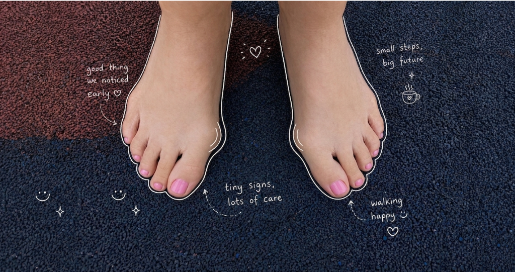
Juvenile hallux valgus, often called a juvenile bunion, is a condition where the big toe gradually angles toward the second toe. A bump may appear on the inside of the foot at the base of the big toe. In some children, it is mainly a cosmetic concern. In others, it may cause pain, shoe irritation, difficulty with sports, or progressive change in foot shape.
For parents, the most important point is this: juvenile hallux valgus is not always caused by “bad shoes.” Shoes can make symptoms worse, but in many children the condition is linked to inherited foot structure, joint flexibility, flat feet, first-ray instability, bone shape, and the way the foot functions during walking or running.
Because juvenile hallux valgus is multifactorial, treatment should not be one-size-fits-all. The goal is to understand why this child’s bunion is developing, whether it is progressing, whether it is painful, and which mechanical factors can realistically be improved.
What is juvenile hallux valgus?
In adults, bunions are often associated with years of footwear pressure, joint changes, and progressive deformity. In children and teenagers, the condition is more often influenced by growth, inherited anatomy, ligamentous laxity, flatfoot mechanics, and the shape of the first metatarsal and medial cuneiform bones. A major review of paediatric hallux valgus describes the condition as challenging because management must account for growth, symptoms, deformity severity, recurrence risk, and the limitations of conservative and surgical care.
Juvenile hallux valgus may affect one foot or both feet. It is commonly noticed during late childhood or adolescence, but in some children the shape is visible earlier.
Common signs parents may notice
- The big toe leaning toward the second toe
- A bump on the inside of the forefoot
- Redness or irritation from shoes
- Pain after sport or prolonged walking
- Difficulty fitting school shoes, football boots, ballet shoes, or narrow casual shoes
- The second toe being pushed or crowded
- Flattening of the arch or the foot rolling inward
- A strong family history of bunions
Some children have a visible deformity but no pain. Others have mild-looking deformity but significant discomfort. This is why assessment should not rely on appearance alone.
Why does juvenile hallux valgus happen?
1. Family history and inherited foot structure
Many children with juvenile hallux valgus have a parent or close relative with bunions. This does not mean the child will definitely need surgery, but it does suggest an inherited tendency in foot structure and joint mechanics.
Inherited factors may include:
- A wider forefoot
- A more flexible medial column
- Increased first-ray mobility
- Ligamentous laxity
- Flatfoot tendency
- Metatarsus primus varus
- Abnormal angulation of the first metatarsal head
This is one reason parents should avoid blaming the child’s shoes alone. Shoes may irritate the bunion, but they may not be the original cause.
2. The shape of the first metatarsal and medial cuneiform
Research shows that juvenile hallux valgus is often related to the shape and alignment of bones in the medial column of the foot. Kaiser et al. studied standing radiographs of juvenile hallux valgus feet and found that deformity was highly correlated with morphology of the first metatarsal and medial cuneiform. Severe cases were associated with increased bony deformity and increased incongruity of the first metatarsophalangeal joint.
This matters because a child’s bunion may not simply be a soft-tissue problem. If the joint surface itself is angled, the big toe may follow that angled surface into valgus. This helps explain why some juvenile bunions progress despite reasonable shoes and conservative care.
3. Distal metatarsal articular angle
The distal metatarsal articular angle, or DMAA, describes the orientation of the joint surface at the head of the first metatarsal. In juvenile hallux valgus, the DMAA can be increased. When the joint surface faces laterally, the big toe may sit in valgus even when the joint is still relatively congruent.
This is one reason radiographs may sometimes be needed, especially if surgery is being considered. External measurements can show the visible angle of the big toe, but they cannot show DMAA, sesamoid position, joint congruency, or growth plate status.
4. Flat feet and excessive pronation
Many children with juvenile hallux valgus also have flexible flat feet. When the foot rolls inward excessively, the medial side of the foot may become less stable. This can affect the first ray and the way the big toe loads during push-off.
However, the relationship is not simple. Flat feet do not automatically cause bunions, and not every child with juvenile hallux valgus has flat feet. The more accurate view is that flatfoot mechanics may be one contributing factor in some children.
This is why biomechanical assessment is important. The question is not simply, “Does the child have a bunion?” The better question is, “What is this child’s foot doing during gait, and is that movement contributing to symptoms or progression?”
5. Ligamentous laxity and hypermobility
Some children have generally flexible joints. This may allow the forefoot to splay and the first ray to move excessively during walking or sport. In a child with a strong family history and flexible feet, ligamentous laxity may increase the risk of progression.
Clinical assessment should therefore include more than the big toe. The clinician should assess the arch, heel position, subtalar joint movement, first-ray mobility, calf tightness, knee alignment, and general joint flexibility.
6. Footwear pressure
Narrow shoes, pointed shoes, tight school shoes, ballet shoes, and slim sports footwear can worsen symptoms by compressing the big toe toward the second toe. Footwear may also rub the medial bump and cause redness or pain.
Footwear is rarely the only cause of juvenile hallux valgus, but it can be an important aggravating factor. A child with a structural tendency toward hallux valgus may become symptomatic sooner if footwear is narrow, shallow, or unstable.
Does juvenile hallux valgus get worse?
That does not mean every child’s bunion will worsen at the same rate. Progression depends on several factors, including:
- Age
- Growth stage
- Family history
- Severity at presentation
- Footwear habits
- Flatfoot mechanics
- Joint laxity
- First-ray instability
- Bony morphology
- Whether the first MTP joint remains congruent
- Activity level and sport demands
This is why regular monitoring can be helpful. The goal is not to X-ray every child repeatedly. The goal is to monitor the child intelligently, using clinical and biomechanical tools, and reserve radiographs for situations where they are clinically necessary.
When should parents seek assessment?
- The bunion is painful
- The deformity appears to be worsening
- The child avoids sport or activity because of foot pain
- Shoes rub or become difficult to fit
- The second toe is being pushed or lifted
- There is a strong family history of severe bunions
- The child also has flat feet, knee pain, ankle pain, or frequent fatigue
- One foot is much worse than the other
- The child is very young and already has a noticeable deformity
Early assessment does not mean early surgery. In most cases, the first step is careful examination, footwear advice, functional monitoring, and conservative management.
How juvenile hallux valgus is assessed
Clinical examination
The clinician should assess:
- Hallux valgus angle
- Flexibility of the deformity
- First MTP joint range of motion
- Pain location
- Medial bump irritation
- Lesser toe crowding
- Arch height
- Heel valgus
- First-ray mobility
- Joint laxity
- Calf tightness
- Walking and running mechanics
- Footwear fit
The examination should also clarify whether the main issue is pain, progression, footwear difficulty, cosmetic concern, or functional limitation.
Biomechanical assessment
Biomechanical assessment helps determine how the foot behaves under load. This is especially important in children because the deformity may be influenced by gait mechanics, not simply static foot shape.
For example, two children may have a similar-looking bunion. One may have a stable arch, good first-ray function, and mainly shoe irritation. Another may have flexible flat feet, delayed resupination, excessive medial column collapse, and poor propulsion through the big toe. These children should not receive the same plan.
Are X-rays always needed?
Weight-bearing radiographs remain the reference standard for measuring structural hallux valgus severity and assessing deeper features such as intermetatarsal angle, DMAA, sesamoid position, joint congruency, and growth plates. Janssen et al. noted that radiographic measurements are generally considered the most reproducible and accurate method for hallux valgus angle assessment.
However, repeated radiographs are not always necessary in a growing child, especially when the child is being monitored conservatively and there is no immediate surgical question. In healthcare, radiation exposure should be justified and minimized where reasonable. This is especially relevant in children, who may be monitored over several years.
A balanced approach is best:
X-rays may be appropriate when:
- The deformity is severe
- The bunion is painful and progressive
- Surgery is being considered
- The diagnosis is uncertain
- There is asymmetry, stiffness, trauma history, or unusual presentation
- Structural angles such as IMA or DMAA need to be measured
X-rays may not be needed repeatedly when:
- The deformity is mild
- The child is being monitored conservatively
- Symptoms are improving
- There is no surgical decision to make
- Clinical and digital measurements are stable
Can bunion angles be measured without X-rays?
Non-radiographic methods include:
- Clinical angle measurement
- Goniometer measurement
- Digital photographic measurement
- Smartphone-based measurement
- Visual grading scales such as the Manchester Scale
- Plantar pressure and functional assessment
Digital photographic measurement has shown promising reliability and validity in hallux valgus assessment. Nix et al. concluded that digital photographic measurements of hallux valgus angle are reliable and have acceptable validity compared with weight-bearing radiographs. Cakar et al. also found that hallux valgus angle measurement on smartphone digital photographs showed strong reliability and validity.
Clinical goniometer measurement has also been studied. Janssen et al. compared hallux valgus angles assessed using radiographs, clinical examination, and computerised plantar pressure measurement; their findings support the idea that clinical measurement can be useful, although radiographs remain the reference standard. Choung et al. specifically examined the reliability and validity of goniometer measurement compared with radiographs, but later summaries report that goniometer measurement may have unacceptable validity when used as a radiographic substitute.
The practical conclusion is straightforward:
Digital and clinical angle measurements are useful for monitoring visible progression, but they do not replace X-rays when deeper structural assessment is required.
What is the role of plantar pressure analysis?
Plantar pressure analysis measures how force is distributed under the foot during walking. It does not simply show what the foot looks like; it shows how the foot loads.
This can be useful in juvenile hallux valgus because the big toe plays an important role during propulsion. Hallux valgus can alter loading under the hallux and medial forefoot. Studies have reported altered plantar loading patterns in hallux valgus feet, including changes in load beneath the hallux and first metatarsal region.
In children, plantar pressure data can help answer questions such as:
- Is the child loading the big toe properly during push-off?
- Is there excessive pressure under the medial forefoot?
- Is the foot rolling inward excessively?
- Is there asymmetry between the left and right foot?
- Is the child compensating because of pain?
- Is the orthotic or footwear improving loading?
Plantar pressure analysis should not be used alone to diagnose hallux valgus severity. It is best used as part of a broader biomechanical assessment.
Conservative treatment: what can help?
Most children with juvenile hallux valgus start with conservative care. The aim is not to promise that the bunion will disappear. The aim is to reduce pain, improve function, reduce irritation, address contributing mechanics, and monitor progression.
1. Footwear modification
Footwear advice is often the first and most important step.
Useful shoe features include:
- Wide toe box
- Adequate depth
- Firm heel counter
- Stable midsole
- Removable insole if orthoses are needed
- Avoidance of pointed or narrow forefoot shapes
- Avoidance of shoes that force the big toe inward
- Appropriate width for school shoes and sports shoes
Parents should check the shoes the child actually wears most often. A child may attend clinic in good shoes but spend most of the week in narrow school shoes, football boots, or dance shoes.
2. Orthoses
Foot orthoses may help when juvenile hallux valgus is associated with:
- Flexible flat feet
- Excessive pronation
- Medial column instability
- First-ray hypermobility
- Fatigue during walking
- Pain during sport
- Abnormal pressure distribution
- Poor shoe mechanics
However, orthoses should be explained honestly. They may improve function and reduce symptoms, but they do not reliably straighten a juvenile bunion.
A controlled prospective trial by Kilmartin, Barrington and Wallace studied foot orthoses in children with juvenile hallux valgus. The authors found that the metatarsophalangeal joint angle increased in both treated and untreated groups, and hallux valgus developed in some previously unaffected feet despite orthotic use.
This does not mean orthoses have no role. It means orthoses should not be presented as a guaranteed bunion-correction device. Their role is more appropriate when they are used to improve foot mechanics, reduce symptoms, support the medial column, and manage associated flatfoot function.
3. Toe spacers and splints
Toe spacers may reduce pressure between the first and second toes and may improve comfort in shoes. Night splints may temporarily position the toe more comfortably. These devices may be helpful for symptoms, especially in flexible deformities, but they are unlikely to permanently correct a structural juvenile bunion.
4. Exercises and strengthening
Exercises may be useful when the child has poor foot control, weak intrinsic muscles, calf tightness, or excessive pronation. Exercises may include:
- Short-foot activation
- Toe control exercises
- Calf stretching
- Balance work
- Strengthening of the foot and ankle stabilizers
- Gait retraining in selected cases
Exercises should be viewed as one part of a wider plan, not as a stand-alone cure.
5. Activity and sport modification
Most children should remain active. The aim is not to stop sport unnecessarily. However, certain shoes and activities may aggravate symptoms. Football boots, ballet shoes, dance footwear, and narrow court shoes may need specific advice.
If pain is activity-related, management may include temporary load modification, shoe changes, padding, orthoses, or sport-specific footwear recommendations.
When is surgery considered?
Surgery is usually not the first treatment for juvenile hallux valgus. It may be considered when there is persistent pain, progressive deformity, difficulty with shoes, or functional limitation despite appropriate conservative care.
Surgical decision-making in adolescents is complex because recurrence has historically been a concern. A systematic review by Harb et al. found that contemporary surgical interventions for adolescent hallux valgus can show good clinical and radiological outcomes with high patient satisfaction, but the authors also emphasized that adolescent hallux valgus management remains controversial.
Surgery should be planned according to the deformity, not merely the appearance of the bunion. Important surgical considerations include:
- Skeletal maturity
- Hallux valgus angle
- Intermetatarsal angle
- DMAA
- Sesamoid position
- MTP joint congruity
- First-ray instability
- Flatfoot contribution
- Ligamentous laxity
- Severity of symptoms
For many children, the best approach is careful conservative care and monitoring until skeletal maturity, unless symptoms or progression justify earlier specialist referral.
How Family Podiatry Centre monitors juvenile hallux valgus
At Family Podiatry Centre, our approach is based on a simple principle:
We do not treat every juvenile bunion the same way, because juvenile hallux valgus does not have one single cause.
Some children mainly need footwear advice and monitoring. Some need orthotic support because flatfoot mechanics or excessive pronation are contributing to symptoms. Some need sport-specific shoe advice. Some need digital angle monitoring. Some need referral for radiographs or surgical opinion if the deformity is severe, painful, or progressive.
1. We begin with a full clinical assessment
We assess:
- The visible bunion angle
- Pain location
- Shoe irritation
- Flexibility of the big toe joint
- First-ray mobility
- Arch height
- Heel position
- Flatfoot mechanics
- General joint flexibility
- Calf tightness
- Toe crowding
- Family history
- Sport and footwear habits
This helps us identify whether the bunion is mainly a shoe-pressure problem, a flatfoot-related mechanical issue, a structural inherited deformity, or a more complex combination.
2. We use emed plantar pressure analysis
Family Podiatry Centre uses emed plantar pressure analysis to assess how the child’s foot loads during gait. This allows us to examine foot function dynamically, rather than relying only on static appearance.
The emed system helps us evaluate:
- Pressure under the hallux
- Medial forefoot loading
- Timing of pressure transfer
- Left-right asymmetry
- Excessive medial loading
- Compensation patterns
- Whether orthoses or shoe changes improve loading
This is particularly useful because juvenile hallux valgus is not only about the big toe angle. It is also about how the foot behaves under load.
3. We use video gait analysis
Video gait analysis allows us to observe:
- Foot progression angle
- Heel position
- Arch collapse
- Timing of pronation and resupination
- Knee and hip compensation
- Propulsion pattern
- Running or sport-specific mechanics when relevant
This helps us decide whether the child’s hallux valgus is part of a wider lower-limb movement pattern.
4. We use digital bunion angle measurement
Rather than repeatedly sending a child for X-rays just to check whether the visible angle has changed, we can use digital photographic bunion angle measurement as a clinical monitoring tool.
This is useful for:
- Baseline documentation
- Follow-up comparison
- Monitoring visible progression
- Parent education
- Reducing unnecessary repeated radiation exposure
Digital photographic measurement is supported by studies showing good reliability and acceptable validity compared with radiographs, although it does not replace X-rays when structural assessment is required.
5. We also use clinical angle measurement
Clinical angle measurement can help track the visible deformity over time. It is not a perfect substitute for radiographs, and we do not present it as one. However, it can be useful when combined with photographs, gait analysis, pressure analysis, symptoms, and footwear assessment.
This layered approach allows us to monitor the child while avoiding unnecessary repeated radiographs.
6. We refer for X-rays when they are clinically necessary
We do not avoid X-rays when they are needed. X-rays are important when deeper structural information is required, especially in more severe or progressive cases.
Radiographs may be recommended if:
- The deformity is worsening rapidly
- Pain is persistent
- The joint is stiff or incongruent
- Surgical opinion may be needed
- There is significant asymmetry
- We need to assess IMA, DMAA, sesamoid position, or growth plate status
Our approach is not “no X-rays.” It is appropriate X-rays when clinically justified, and non-radiographic monitoring when that is sufficient.
7. We tailor treatment to the child
Because juvenile hallux valgus is multifactorial, treatment may include several elements:
- Footwear changes
- Orthoses where biomechanics justify them
- Toe spacers for comfort
- Padding for shoe irritation
- Strengthening and mobility exercises
- Calf stretching
- Sport-specific footwear advice
- Digital angle monitoring
- emed pressure reassessment
- Video gait reassessment
- Referral for imaging or surgical opinion when appropriate
This allows us to manage the child’s symptoms, monitor progression, and address the mechanical factors that may be contributing to the problem.
Key message for parents
A juvenile bunion should not be ignored, but it also should not be over-treated.
The best first step is a careful assessment that asks:
- Is it painful?
- Is it progressing?
- Is the child’s foot function contributing to it?
- Are shoes making it worse?
- Is there flatfoot or excessive pronation?
- Is there a strong family history?
- Can we monitor it safely without repeated X-rays?
- Does the child need conservative care, imaging, or referral?
At Family Podiatry Centre, we use clinical assessment, emed plantar pressure analysis, video gait analysis, digital bunion angle measurement, and clinical angle measurement to monitor juvenile hallux valgus in a child-friendly, function-focused way. Radiographs remain important when structural assessment is needed, but repeated X-rays are not always necessary for routine monitoring.
Book An Appointment
Disclaimer: The word "treatment" in this article refers to the care and management of a patient’s health to prevent, cure, or improve a condition. Treatment results vary and do not necessarily indicate a cure. This article is for informational and educational purposes only and does not constitute medical advice.
About the Author
Mark B. Reyneker, BTech (Podiatry), MSc (Palaeontology) is a podiatrist and Founder & Clinical Director of Family Podiatry Centre, with more than 25 years of clinical experience across South Africa, Malaysia and Singapore. His clinical interests include foot and lower-limb pain, gait and biomechanics, sports-related foot conditions, orthotic therapy and footwear.
Alongside his clinical practice, Mark conducts research into human gait and foot biomechanics. His MSc research at the University of the Witwatersrand investigated human propulsion and the structural properties of the metatarsals. He is also the inventor of A Foot Orthotic, an orthotic technology developed through an international patent family.
References
2. Choung, S.D., Kang, S.Y., Kim, M.H. and Weon, J.H. (2013) ‘Reliability and validity of the goniometer for hallux valgus angle measurement’, Physical Therapy Korea, 20(2), pp. 46–51.
3. Galica, A.M., Hagedorn, T.J., Dufour, A.B., Riskowski, J.L., Hillstrom, H.J., Casey, V.A. and Hannan, M.T. (2013) ‘Hallux valgus and plantar pressure loading: the Framingham foot study’, Journal of Foot and Ankle Research, 6, article 42.
4. Garrow, A.P., Papageorgiou, A.C., Silman, A.J., Thomas, E., Jayson, M.I.V. and Macfarlane, G.J. (2001) ‘The grading of hallux valgus: the Manchester Scale’, Journal of the American Podiatric Medical Association, 91(2), pp. 74–78.
5. Harb, Z., Kokkinakis, M., Ismail, H. and Spence, G. (2015) ‘Adolescent hallux valgus: a systematic review of outcomes following surgery’, Journal of Children’s Orthopaedics, 9(2), pp. 105–112.
6. Janssen, D.M.C., Sanders, A.P., Guldemond, N.A., Hermus, J., Walenkamp, G.H.I.M. and van Rhijn, L.W. (2014) ‘A comparison of hallux valgus angles assessed with computerised plantar pressure measurements, clinical examination and radiography in patients with diabetes’, Journal of Foot and Ankle Research, 7, article 33.
7. Kaiser, P., Livingston, K., Miller, P.E., May, C. and Mahan, S. (2018) ‘Radiographic evaluation of first metatarsal and medial cuneiform morphology in juvenile hallux valgus’, Foot & Ankle International, 39(10), pp. 1223–1228.
8. Kilmartin, T.E., Barrington, R.L. and Wallace, W.A. (1994) ‘A controlled prospective trial of a foot orthosis for juvenile hallux valgus’, Journal of Bone and Joint Surgery. British Volume, 76-B(2), pp. 210–214.
9. Lee, K.M., Ahn, S., Chung, C.Y., Sung, K.H., Park, M.S. and Lee, S.Y. (2012) ‘Reliability and relationship of radiographic measurements in hallux valgus’, Clinical Orthopaedics and Related Research, 470, pp. 2613–2621.
10. Li, G., Shen, J., Smith, E. and Patel, C. (2022) ‘Development of a manual measurement device for measuring hallux valgus angle in patients with hallux valgus’, International Journal of Environmental Research and Public Health, 19(15), article 9108.
11. Menz, H.B., Fotoohabadi, M.R., Wee, E. and Spink, M.J. (2010) ‘Validity of self-assessment of hallux valgus using the Manchester scale’, BMC Musculoskeletal Disorders, 11, article 215.
12. Nix, S., Russell, T., Vicenzino, B. and Smith, M. (2012) ‘Validity and reliability of hallux valgus angle measured on digital photographs’, Journal of Orthopaedic & Sports Physical Therapy, 42(7), pp. 642–648.
13. Seidenstein, A.H., Torrez, T.W., Mahan, S.T. and Toy, J.O. (2021) ‘Pediatric hallux valgus: an overview of history, examination, conservative, and surgical management’, Journal of Children’s Orthopaedics, 15(5), pp. 457–463.
14. Sung, K.H., Kwon, S.S., Park, M.S., Lee, K.M., Ahn, J. and Lee, S.Y. (2019) ‘Natural progression of radiographic indices in juvenile hallux valgus deformity’, Foot and Ankle Surgery, 25(3), pp. 378–382.
15. Wong, D.W.C., Wang, Y., Zhang, M. and Leung, A.K.L. (2025) ‘Does hallux valgus impair medial forefoot loading? A meta-analysis’, Journal of Clinical Medicine / related biomechanics literature.
Frequently Asked Questions
Yes. Children and teenagers can develop bunions, known medically as juvenile hallux valgus. This happens when the big toe gradually angles toward the second toe, sometimes creating a visible bump on the inside of the foot. In some children it causes little pain, while in others it may lead to shoe irritation, sport-related discomfort, or progressive change in foot shape.
Juvenile hallux valgus is usually multifactorial. It is not always caused by tight or “bad” shoes. Common contributing factors include inherited foot structure, family history, flexible flat feet, excessive pronation, ligamentous laxity, first-ray instability, bone shape, and the way the foot loads during walking or running. Shoes may aggravate symptoms, but they are rarely the only cause.
Parents should consider a podiatry assessment if the bunion is painful, appears to be getting worse, causes shoe-fitting problems, affects sport, pushes the second toe, or is associated with flat feet, ankle pain, knee pain, or fatigue. A strong family history of severe bunions is another reason to monitor the condition earlier.
No. X-rays are not always needed at every visit. Weight-bearing X-rays can be useful when the deformity is severe, painful, progressive, unusual, or when surgery is being considered. For many children, clinical assessment, digital angle monitoring, gait analysis, and plantar pressure analysis can help monitor visible progression without repeated radiographs.
Conservative treatment may reduce pain, improve foot function, reduce shoe irritation, and help manage contributing mechanics, but it should not be presented as a guaranteed way to permanently straighten a juvenile bunion. Footwear changes, orthoses, toe spacers, exercises, strengthening, and activity modification may all help depending on the child’s symptoms, foot posture, and gait pattern.