Published: 12 July 2026 | Last reviewed: 13 July 2026
Hallux valgus is more than the visible bump commonly called a bunion. It is a progressive change in the alignment and function of the big-toe joint. This article explains why hallux valgus develops, why early diagnosis matters and what current research tells us about reducing pain—and, in selected mild-to-moderate cases, improving the angle—without surgery.
Introduction
Hallux valgus is one of the most common structural problems affecting the forefoot. It is often described simply as a “bunion”, but the two terms do not mean exactly the same thing.
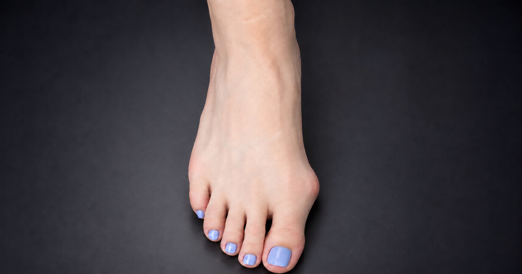
A bunion is the visible prominence that develops on the inner side of the big-toe joint. Hallux valgus is the underlying change in the alignment of the big toe, first metatarsal and surrounding soft tissues. In other words, the bunion is the visible sign; hallux valgus is the structural and mechanical process that produces it.
This distinction is important because treating only the visible bump may miss the deeper problem. Hallux valgus is not simply a lump of extra bone. It is a progressive change in joint alignment, tendon balance, soft-tissue tension, sesamoid position and forefoot loading. The condition is common. A systematic review by Nix, Smith and Vicenzino (2010) estimated that hallux valgus affects approximately 23% of adults aged 18–65 years and approximately 35.7% of people over 65 years. A more recent global systematic review estimated the pooled prevalence at approximately 19%, with higher rates in women and older adults (Cai et al., 2023).
For many people, the main concern is pain, footwear difficulty or the cosmetic appearance of the foot. For others, the bunion is part of a wider forefoot problem involving the second toe, central forefoot overload, callus formation or altered walking mechanics.
This article focuses on non-surgical management. It explains why hallux valgus develops, why early identification matters and what the research says about conservative treatment—particularly the important difference between reducing pain and reducing the actual hallux valgus angle.
What is the difference between a bunion and hallux valgus?
The word hallux means big toe. Valgus means that the toe deviates away from the midline of the body, towards the smaller toes.
Hallux valgus therefore describes the big toe drifting towards the second toe. However, the condition involves more than the toe leaning sideways.
As hallux valgus develops:
- the big toe moves towards the second toe;
- the first metatarsal moves towards the inner side of the foot;
- the joint capsule becomes stretched on one side and tight on the other;
- the tendons around the joint begin to pull from less favourable positions;
- the sesamoid bones beneath the joint become displaced relative to the metatarsal head;
- the big toe may rotate, causing the toenail to face slightly inwards;
- the second toe may become crowded, lifted or unstable.
The bunion is the visible prominence created by these deeper structural changes. It may include the prominent first metatarsal head, thickened soft tissue, local inflammation and sometimes secondary bone remodelling.
A bunion is therefore not the complete diagnosis. It is one feature of hallux valgus.
A bunion is not simply a growth of bone
A common misconception is that a bunion is a new piece of bone growing from the side of the foot. Although the bone and soft tissues can remodel over time, the main reason the bump appears is that the first metatarsal has moved outwards while the big toe has moved in the opposite direction.
This explains why hallux valgus cannot be fully understood by looking only at the size of the bump. The alignment of the whole first ray—the first metatarsal, big-toe joint, sesamoids and surrounding soft tissues—must be considered.
Once the big toe and first metatarsal begin to move away from their normal positions, the joint can become mechanically disadvantaged. Some structures that originally helped stabilise the big toe may begin to contribute to further deformity. The medial joint capsule stretches, the lateral structures tighten and the big toe may gradually become less reducible.
This is one reason hallux valgus may become self-reinforcing. The more the joint changes, the less efficiently its normal stabilising structures can control it.
What causes hallux valgus?
Hallux valgus does not have one single cause. It is best understood as a multifactorial condition. Genetics, foot shape, joint mechanics, connective-tissue properties, age, footwear and loading patterns may all contribute.
Two people may develop similar-looking bunions for different reasons. One person may have strong inherited susceptibility. Another may have a relatively flexible first ray, narrow footwear exposure, forefoot overload or a combination of these factors.
Genetic and familial factors
Family history is one of the strongest associations. Piqué-Vidal, Solé and Antich (2007) reported a strong hereditary pattern in a pedigree study of 350 patients with hallux valgus. Twin and genetic studies also support a heritable component, although hallux valgus is not caused by one single gene (Lee et al., 2014; Hsu et al., 2015).
What may be inherited is not necessarily the bunion itself, but a combination of features that increase susceptibility. These may include:
- the shape of the first metatarsal;
- the orientation of the joint surface;
- ligamentous laxity;
- foot width;
- metatarsal length pattern;
- first-ray mobility;
- connective-tissue characteristics;
- the way the foot loads during walking.
This helps explain why bunions often “run in families” but still vary in severity between relatives.
Foot structure and first-ray mechanics
The first ray plays a key role in forefoot stability and propulsion. During walking, the big toe should help stabilise the medial forefoot as the body moves forwards.
If the first ray is unstable, poorly aligned or functioning from a mechanically disadvantaged position, the big-toe joint may be exposed to abnormal forces. Over time, this can contribute to progressive deviation of the hallux.
However, first-ray mobility should not be oversimplified. Some people with hallux valgus have increased first-ray mobility, but it is not always clear whether this is a cause or a consequence of the deformity. Doty and Coughlin (2013) emphasised that the relationship between first-ray hypermobility and hallux valgus remains complex.
Foot posture
Hallux valgus is sometimes blamed entirely on flat feet or pronation. This is too simplistic. A pronated foot posture may alter loading through the medial column and big-toe joint in some people. However, many people with flatter feet never develop hallux valgus, and many people with hallux valgus do not have markedly flat feet.
Foot posture should therefore be considered as one possible contributor rather than a universal cause. The more important question is how the foot behaves during standing, walking and propulsion.
Footwear
Narrow footwear can crowd the toes and place prolonged pressure across the forefoot. Elevated heels may increase load through the front of the foot and push the toes into the narrowest part of the shoe. Footwear should not be blamed as the only cause of hallux valgus, but it can be an important environmental factor. In a susceptible foot, footwear may increase symptoms, accelerate irritation or maintain the big toe in a deviated position for long periods.
A helpful way to understand footwear is as a modifier. It may not create hallux valgus by itself in every person, but it can aggravate a structural tendency that already exists.
Age and sex
Hallux valgus becomes more common with age and is more common in women. The higher prevalence in women is often attributed to footwear, but this is unlikely to be the only explanation.
Other possible contributors include differences in foot shape, ligamentous laxity, connective-tissue properties, hormonal influences and lifetime loading patterns. Age may also reduce the capacity of the soft tissues and muscles to stabilise the medial forefoot.
Importantly, hallux valgus is not only a condition of older adults. It can appear in childhood, adolescence or early adulthood, especially where there is strong family history or marked structural susceptibility.
Medical and systemic contributors
Hallux valgus may also be associated with inflammatory arthritis, neuromuscular conditions, connective-tissue disorders and previous trauma. These cases require careful assessment because the underlying condition may affect prognosis and treatment planning.
How hallux valgus affects the rest of the forefoot
The big toe is not an isolated structure. It is part of the forefoot loading system.
When the big toe and first metatarsal lose their normal alignment, pressure may shift towards the lesser metatarsals. This can contribute to pain beneath the central forefoot, callus formation, second-toe instability, plantar-plate overload, hammertoe development or crossover-toe deformity.
This is why a patient may present with pain under the second or third metatarsal rather than pain directly over the bunion. The visible bunion may be only one part of a wider mechanical pattern.
Hallux valgus can therefore affect:
- footwear comfort;
- walking efficiency;
- forefoot pressure distribution;
- second-toe stability;
- balance;
- tolerance for exercise;
- confidence during push-off.
The aim of treatment should not be limited to making the bump less painful. The broader goal is to improve the way the forefoot functions.
Pain and deformity do not always match
One of the most important clinical points is that pain and deformity severity do not always correlate. A patient may have a large bunion with surprisingly little pain. Another may have a mild or moderate deformity that is very painful. Pain may arise from pressure over the medial prominence, inflammation of the joint capsule or bursa, irritation within the joint, transfer loading under the lesser metatarsals or associated second-toe instability.
This helps explain why non-surgical treatment can be useful even when the angle does not change dramatically. If the painful tissues can be unloaded or irritated less often, the patient may experience meaningful improvement despite the bunion still being visible. It also explains why surgery should not be recommended purely because a bunion looks large. Surgery is usually considered when pain, function or progressive deformity creates a significant problem—not simply because the foot does not look straight.
Is hallux valgus always progressive?
Hallux valgus is often progressive, but not every case progresses at the same speed.
Some deformities remain stable for many years. Others worsen gradually. A smaller number may progress more quickly, particularly where the deformity is flexible early on but exposed to continued mechanical stress, or where there is strong inherited susceptibility.
Progression may involve:
- increasing big-toe deviation;
- increasing bunion prominence;
- rotation of the big toe;
- worsening crowding of the second toe;
- reduced joint mobility;
- increasing transfer pressure under the forefoot;
- increasing difficulty fitting shoes.
Predicting progression in an individual patient is difficult. This is why monitoring is valuable, especially when hallux valgus is noticed early.
Why early identification matters
Early identification is one of the most important parts of non-surgical care.
This does not mean that every slight deviation of the big toe requires intensive treatment. It means that a developing hallux valgus pattern should be recognised before the deformity becomes rigid, painful or associated with secondary problems.
In an early or moderate case, the big toe may still be flexible. The joint may remain reasonably congruent. The second toe may not yet be unstable. The sesamoids may not be severely displaced. The soft tissues may not yet have developed fixed shortening or stretching. This creates a more favourable opportunity for non-surgical care.
Early assessment allows the clinician to determine:
- whether the deformity is flexible or fixed;
- whether the joint remains healthy;
- whether the toe is beginning to rotate;
- whether the second toe is becoming crowded or unstable;
- whether pressure is shifting towards the lesser metatarsals;
- whether the problem is progressing;
- whether the patient has modifiable mechanical contributors.
Early treatment cannot guarantee that hallux valgus will stop progressing. However, a flexible mild or moderate deformity offers a better opportunity than a severe, rigid deformity with advanced joint change.
Early signs of hallux valgus
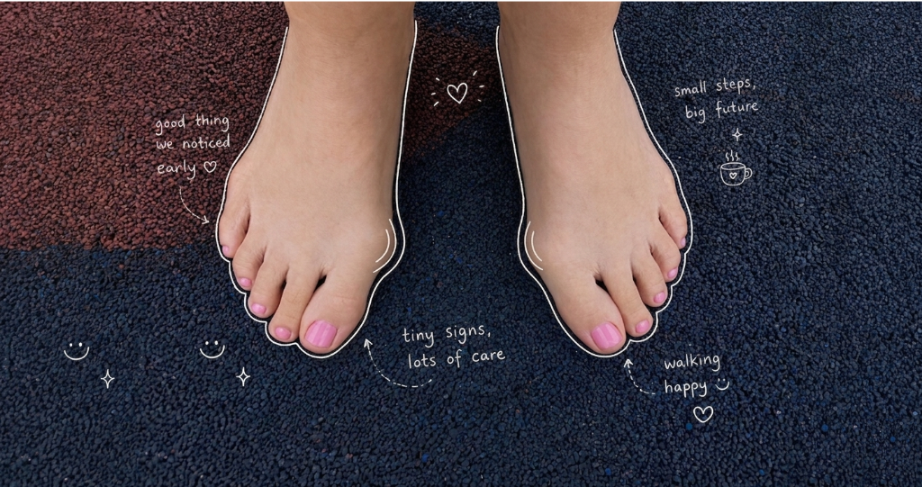
The first sign is not always pain. Early features may include:
- the big toe beginning to lean towards the second toe;
- a widening forefoot;
- redness over the inner side of the big-toe joint after wearing shoes;
- intermittent tenderness around the big-toe joint;
- new difficulty fitting previously comfortable shoes;
- rotation of the big toe or toenail;
- reduced space between the first and second toes;
- callus beneath the central forefoot;
- the second toe beginning to lift, drift or overlap;
- a strong family history combined with visible toe deviation.
Pain should not be the only reason to seek assessment. By the time pain is constant, the deformity may already be more established.
How hallux valgus is assessed
A proper assessment should examine more than the appearance of the bunion.
The clinician may assess:
- the angle of the big toe;
- the position of the first metatarsal;
- whether the deformity is flexible or fixed;
- the movement of the first metatarsophalangeal joint;
- whether the big toe is rotating;
- the position and stability of the second toe;
- callus and pressure patterns;
- foot posture;
- first-ray function;
- walking mechanics;
- pain location;
- family history;
- footwear history;
- whether the deformity appears to be changing over time.
Weight-bearing radiographs may be used when more detailed structural assessment is needed. They can measure the hallux valgus angle, intermetatarsal angle, joint congruency, sesamoid position and signs of arthritis.
Clinical photographs and serial measurements can also be useful, especially for monitoring progression over time.
What does successful non-surgical treatment mean?
Success must be defined carefully.
For some patients, success means less pain. For others, it means walking comfortably, tolerating shoes, exercising again or preventing the second toe from becoming overloaded. In selected mild-to-moderate cases, success may also include measurable improvement in the hallux valgus angle.
Success does not always mean that the adult toe becomes completely straight. This distinction is critical. Conservative care can be very worthwhile even when it does not erase the visible bunion.
Can non-surgical treatment reduce the hallux valgus angle?
It has traditionally been stated that conservative treatment may reduce bunion pain but cannot alter the hallux valgus deformity. That statement is too absolute.
Current research suggests that some people with mild-to-moderate hallux valgus can achieve a measurable reduction in the hallux valgus angle through structured, clinician-directed non-surgical care.
A network meta-analysis by Zhu et al. (2025) included 11 randomised controlled trials involving 401 patients with predominantly mild-to-moderate hallux valgus. The authors found that conservative programmes could reduce the hallux valgus angle, with average reductions of approximately 3.3° for active conservative care and larger estimated reductions in some combined programmes. Pain also improved meaningfully in the better-performing treatment groups.
This supports what is often seen clinically: mild and moderate cases, especially when flexible, may have greater potential for both pain reduction and measurable angular improvement.
Other reviews have reached similar but cautious conclusions. Ying et al. (2021) found that several conservative approaches showed potential for improving hallux valgus angle, intermetatarsal angle and patient symptoms. Hurn et al. (2022) concluded that non-surgical interventions may improve pain and, in some studies, alignment, but emphasised that the available trials were generally small and varied in quality.
The correct conclusion is therefore balanced:
Non-surgical care cannot be promised to permanently straighten every bunion. However, measurable angular improvement is possible in selected mild-to-moderate cases, particularly when the deformity remains flexible and treatment is started early.
Why mild and moderate cases may respond better
A mild or moderate hallux valgus deformity is often more flexible. During examination, the big toe may still be manually reducible towards a better position. The joint surface may remain relatively congruent. The soft tissues may not yet be fixed in their abnormal positions.
At this stage:
- the joint may still have good mobility;
- the soft-tissue imbalance may be less established;
- the sesamoid displacement may be less advanced;
- the big toe may not yet be severely rotated;
- the second toe may not yet be damaged or unstable;
- the forefoot may still respond to mechanical change.
This creates a better opportunity for treatment to influence both symptoms and alignment.
By contrast, once hallux valgus becomes severe, rigid or arthritic, the deformity may be more structurally fixed. In those cases, non-surgical care may still reduce pain and improve function, but large angular correction is less predictable.
Can severe hallux valgus improve without surgery?
Yes, but the goal is usually different.
In severe hallux valgus, the toe may be too rigid or structurally altered to produce major angular correction through conservative care alone. However, pain can still improve.
Nakagawa et al. (2019) followed patients with symptomatic hallux valgus for two years during nonoperative treatment. Pain scores improved significantly and the treatment effect was maintained over 24 months, even though the hallux valgus and intermetatarsal angles did not change during the study period.
This finding is clinically important. It shows that pain reduction and angular correction are not the same outcome.
A severe bunion may become less painful if the irritated tissues are protected, pressure is reduced, joint stress is modified or transfer loading improves. The patient may walk better, tolerate footwear more comfortably and participate more confidently in daily activity, even if the big toe remains visibly deviated.
This is still a meaningful treatment success.
Our clinical observations
In our clinical experience, response to non-surgical care often differs according to the stage and flexibility of the deformity.
In mild and moderate cases, particularly where the big-toe joint remains mobile and the deformity is reducible, we have observed meaningful pain reduction and measurable improvement in the hallux valgus angle.
In more severe cases, substantial angular correction is less predictable. However, these patients may still achieve considerable pain relief and functional improvement despite the visible deformity remaining present.
These observations are consistent with the emerging research. Mild-to-moderate hallux valgus appears to have greater potential for measurable angular improvement, while pain can improve across a wider range of deformity severities.
This should not be interpreted as a guarantee that every bunion can be straightened without surgery. Outcomes depend on the flexibility of the deformity, joint health, duration of the problem, presence of arthritis, degree of rotation, adherence to management and the individual mechanics of the foot.
Why conservative treatment is still worthwhile
A treatment does not have to completely straighten the toe to be valuable.
Pain is influenced by pressure, tissue irritation, inflammation, joint movement and load distribution. Improving these factors can produce meaningful relief even when the visible deformity remains.
Conservative care may be especially appropriate when:
- the deformity is mild or moderate;
- the joint remains flexible;
- symptoms are intermittent or mechanically triggered;
- there is no major joint arthritis;
- the second toe is not yet severely involved;
- the patient wants to avoid or delay surgery;
- surgery would be disproportionate to the symptoms;
- the patient has realistic expectations.
For an early flexible deformity, both pain reduction and angle improvement may be realistic aims.
For an advanced rigid deformity, the more realistic goals may be pain reduction, improved walking tolerance, improved footwear comfort and protection of the lesser toes.
Can non-surgical treatment stop progression?
The current evidence does not prove that conservative care can reliably stop hallux valgus progression in every patient. Most studies are relatively short, include small samples or focus more on pain than long-term structural change.
Our clinical experience (anecdotal evidence), that has tracked multiple patients over several years, has shown very promising results in reducing the angle, the bunion pain or bump size and stopping progression.
However, it is reasonable to say that early, appropriate management may reduce modifiable stress on the joint and may help selected patients maintain function for longer.
The goals are to:
- reduce pain;
- improve function;
- reduce avoidable mechanical stress;
- monitor progression;
- protect the second toe and central forefoot;
- improve activity tolerance;
- delay or avoid surgery where possible;
- identify cases that are deteriorating despite conservative care.
The earlier the problem is identified, the more flexible and responsive the deformity is likely to be.
When non-surgical treatment is less likely to change the angle
Substantial angular correction is less likely when there is:
- severe deformity;
- a rigid big-toe joint;
- marked rotation of the toe;
- established joint arthritis;
- severe sesamoid displacement;
- long-standing soft-tissue imbalance;
- second-toe dislocation or crossover deformity;
- progressive deformity despite sustained care.
Even in these cases, conservative treatment may still reduce pain or improve function. The key is to define success realistically.
When surgery may need to be discussed
This article focuses on non-surgical treatment, but it is important to be honest about the limits of conservative care.
Surgery may need to be discussed when pain remains persistent, footwear difficulty is severe, walking is significantly limited, the second toe is being damaged or the deformity continues to progress despite appropriate conservative management.
However, surgery is not required simply because a bunion is visible. A painless bunion that is stable and not interfering with function does not automatically need an operation.
Common misconceptions about bunions
“A bunion is just extra bone”
Usually, the prominence is mainly the result of the first metatarsal moving outwards and the big toe moving towards the second toe. It is an alignment problem, not just a bone growth.
“All bunions are caused by shoes”
Shoes can contribute, but hallux valgus is usually multifactorial. Genetics, foot structure, soft-tissue characteristics and loading patterns also matter.
“Conservative treatment cannot change the angle”
This is too absolute. Research suggests that selected mild-to-moderate cases can achieve measurable angular improvement. However, established severe deformity is less likely to straighten substantially without surgery.
“If the angle does not change, treatment has failed”
Not necessarily. Pain relief, better walking, improved shoe comfort and reduced forefoot overload are meaningful outcomes.
“A larger bunion must be more painful”
Not always. Pain depends on tissue irritation, pressure, joint health, loading and activity—not only the size of the bunion.
“You should wait until it is very painful before seeking treatment”
Waiting may mean missing the stage when the deformity is most flexible and responsive. Early assessment is often more useful.
Conclusion
Hallux valgus is more than a bunion. The bunion is the visible prominence, while hallux valgus is the underlying structural and functional change affecting the big toe, first metatarsal, sesamoids and surrounding soft tissues.
The condition is multifactorial. Genetics, foot structure, first-ray mechanics, soft-tissue balance, age, footwear and loading patterns may all contribute.
Non-surgical care should not be dismissed as merely temporary symptom relief. Research now supports a more nuanced view. In selected mild-to-moderate cases, conservative care may reduce pain and produce measurable improvement in the hallux valgus angle. In more severe cases, angular correction is less predictable, but pain and function may still improve significantly.
The most important clinical message is that timing matters. Early hallux valgus is more likely to be flexible, reducible and responsive. Waiting until the bunion is severe, rigid or affecting the second toe may reduce the opportunity for meaningful non-surgical change.
A realistic treatment goal is not always to make the foot look perfect. It is to reduce pain, improve function, protect the forefoot and, where possible, improve alignment before the deformity becomes fixed.
Disclaimer: The word "treatment" in this article refers to the care and management of a patient’s health to prevent, cure, or improve a condition. Treatment results vary and do not necessarily indicate a cure. This article is for informational and educational purposes only and does not constitute medical advice.
About the Author
Mark B. Reyneker, BTech (Podiatry), MSc (Palaeontology) is a podiatrist and Founder & Clinical Director of Family Podiatry Centre, with more than 25 years of clinical experience across South Africa, Malaysia and Singapore. His clinical interests include foot and lower-limb pain, gait and biomechanics, sports-related foot conditions, orthotic therapy and footwear.
Alongside his clinical practice, Mark conducts research into human gait and foot biomechanics. His MSc research at the University of the Witwatersrand investigated human propulsion and the structural properties of the metatarsals. He is also the inventor of
A Foot Orthotic, an orthotic technology developed through an international patent family.
References
2.
Cai, Y., Song, Y., He, M., He, W., Zhong, X., Wen, H. and Wei, Q. (2023) ‘Global prevalence and incidence of hallux valgus: a systematic review and meta-analysis’, Journal of Foot and Ankle Research, 16, article 63. doi: 10.1186/s13047-023-00661-9.5.
Fraissler, L., Konrads, C., Hoberg, M., Rudert, M. and Walcher, M. (2016) ‘Treatment of hallux valgus deformity’, EFORT Open Reviews, 1(8), pp. 295–302. doi: 10.1302/2058-5241.1.000005.7. Hecht, P.J. and Lin, T.J. (2014) ‘Hallux valgus’, Medical Clinics of North America, 98(2), pp. 227–232. doi: 10.1016/j.mcna.2013.10.007.
8.
Hsu, Y.H., Liu, Y., Hannan, M.T., Maixner, W., Smith, S.B., Diatchenko, L., Golightly, Y.M., Menz, H.B., Kraus, V.B., Doherty, M., Wilson, A.G. and Jordan, J.M. (2015) ‘Genome-wide association meta-analyses to identify common genetic variants associated with hallux valgus in Caucasian and African Americans’, Journal of Medical Genetics, 52(11), pp. 762–769. doi: 10.1136/jmedgenet-2015-103142.10.
Külünkoğlu, B.A., Akkubak, Y., Çelik, D. and Alkan, A. (2021) ‘A comparison of the effectiveness of splinting, exercise and electrotherapy in women patients with hallux valgus: a randomized clinical trial’, The Foot, 48, article 101828. doi: 10.1016/j.foot.2021.101828.11.
Lee, C.H., Lee, S., Kang, H., Jung, D.E., Song, Y.M., Lee, K., Hwang, J. and Sung, J. (2014) ‘Genetic influences on hallux valgus in Koreans: the Healthy Twin Study’, Twin Research and Human Genetics, 17(2), pp. 121–126. doi: 10.1017/thg.2014.10.12.
Menz, H.B., Marshall, M., Thomas, M.J., Rathod-Mistry, T., Peat, G.M. and Roddy, E. (2023) ‘Incidence and progression of hallux valgus: a prospective cohort study’, Arthritis Care & Research, 75(1), pp. 166–173. doi: 10.1002/acr.24754.13.
Menz, H.B., Lim, P.Q.X., Hurn, S.E., Mickle, K.J., Buldt, A.K., Cotchett, M.P., Roddy, E., Wluka, A.E., Erbas, B., Batra, M. and Munteanu, S.E. (2023) ‘Nonsurgical management of hallux valgus: findings of a randomised pilot and feasibility trial’, Journal of Foot and Ankle Research, 16, article 78. doi: 10.1186/s13047-023-00677-1.14.
Nakagawa, R., Yamaguchi, S., Kimura, S., Sadamasu, A., Yamamoto, Y., Muramatsu, Y., Sato, Y., Akagi, R., Sasho, T. and Ohtori, S. (2019) ‘Efficacy of foot orthoses as nonoperative treatment for hallux valgus: a 2-year follow-up study’, Journal of Orthopaedic Science, 24(3), pp. 526–531. doi: 10.1016/j.jos.2018.11.003.16. Nix, S.E., Vicenzino, B.T. and Smith, M.D. (2012) ‘Foot pain and functional limitation in healthy adults with hallux valgus: a cross-sectional study’, BMC Musculoskeletal Disorders, 13, article 197. doi: 10.1186/1471-2474-13-197.
19.
Torkki, M., Malmivaara, A., Seitsalo, S., Hoikka, V., Laippala, P. and Paavolainen, P. (2001) ‘Surgery versus orthosis versus watchful waiting for hallux valgus: a randomized controlled trial’, JAMA, 285(19), pp. 2474–2480. doi: 10.1001/jama.285.19.2474.21.
Zhu, Z., Liu, Y., Wang, L., Liu, H. and Wang, J. (2025) ‘Effects of exercise combined with external support on hallux valgus angle and pain: a systematic review and network meta-analysis’, Foot and Ankle Surgery, available online 13 September 2025. doi: 10.1016/j.fas.2025.09.004.