When Heel Pain Comes from the Heel Bone
Most people with pain underneath the heel are told that they have plantar fasciitis. In many cases, the plantar fascia is an important part of the problem. However, the plantar fascia is not the only structure capable of producing heel pain.
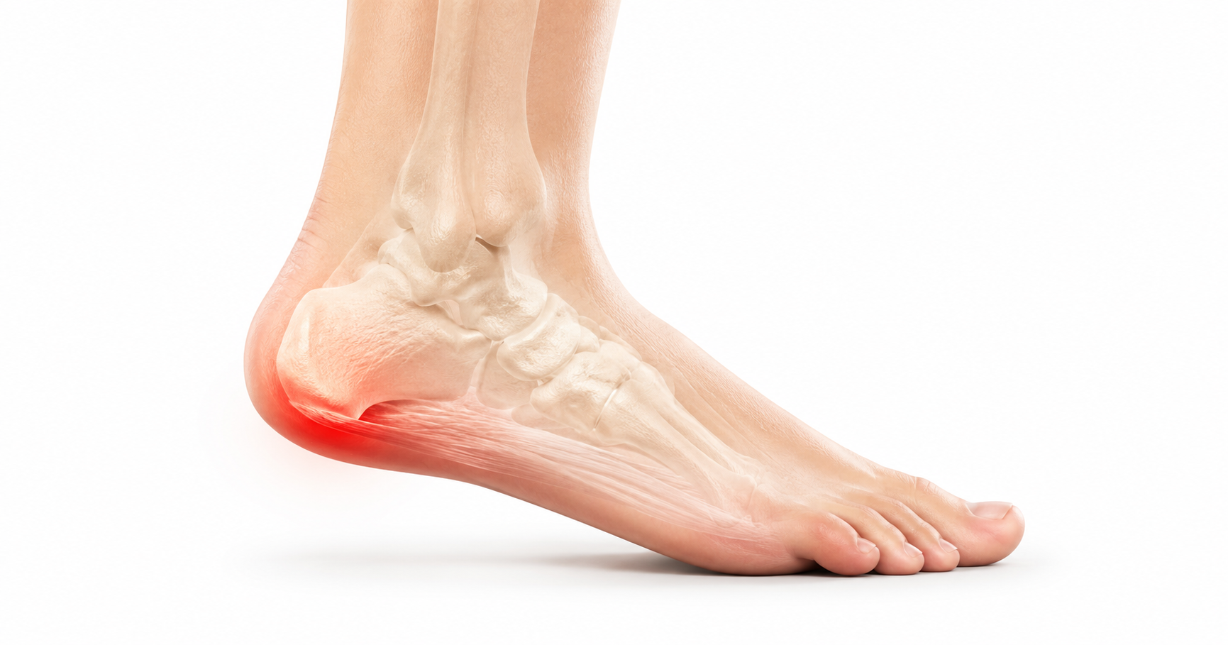
The heel bone itself can also become painful.
The medical name for the heel bone is the calcaneus. Its external surface is covered by a thin, living membrane called the periosteum. This membrane contains blood vessels, bone-forming cells and a rich supply of sensory nerves. It participates in bone growth, adaptation and repair, but it can also become highly sensitive when irritated.
Inflammation or pathological irritation of the periosteum is known as periostitis. When it affects the heel bone, it may be described as calcaneal periostitis.
Calcaneal periostitis can develop when repetitive impact, traction, direct trauma or an underlying disease stimulates the surface of the heel bone. In some patients, the internal part of the calcaneus also becomes involved, producing bone-marrow oedema or a broader bone-stress response.
This means that heel pain does not always fit neatly into a choice between plantar fasciitis and a stress fracture.
The painful process may involve a combination of:
- the plantar fascia;
- the fascia-to-bone attachment;
- the calcaneal periosteum;
- the outer cortical bone;
- the internal trabecular bone and marrow;
- and the cushioning tissues underneath the heel.
Understanding which of these structures is involved is important. A plantar fascial disorder, periosteal irritation and evolving calcaneal stress injury may feel similar, but they do not always require the same degree of protection or the same treatment approach.
What is the periosteum?
The periosteum is a specialised connective-tissue membrane covering most external bone surfaces. It is absent where bones are covered by articular cartilage and has a different structure at certain tendon and ligament attachments.
It has two principal components:
- An outer fibrous layer that provides structural support and carries blood vessels and nerves.
- An inner cellular or cambium layer containing cells that can participate in bone formation, remodelling and repair.
During childhood, the periosteum contributes to bone growth. In adults, it remains important in fracture healing and in the formation of new bone after mechanical or pathological stimulation (Dwek, 2010).
The periosteum is not simply inert wrapping around the bone. It is biologically active and responsive to changes in load.
Why can periostitis be so painful?
Bone is frequently thought of as a solid structure without much sensation. In reality, the periosteum, cortical bone and bone marrow all receive sensory nerve fibres.
The concentration of pain-sensing fibres is greatest in the periosteum, making it particularly sensitive to stretching, pressure, chemical irritation and mechanical disruption. These sensory fibres include thinly myelinated A-delta fibres and unmyelinated C fibres, which transmit sharp and aching pain signals respectively (Oostinga et al., 2020).
This helps explain why a relatively small area of periosteal irritation may produce pronounced local tenderness.
Pain from inside the calcaneus can also occur when bone remodelling, microdamage, increased intraosseous pressure or marrow oedema stimulates nerves within the bone. Consequently, pain described as “heel bone inflammation” may arise from the periosteum, deeper calcaneal bone or both.
What does heel bone inflammation actually mean?
“Heel bone inflammation” is a useful patient description, but it is not one precise pathological diagnosis.
Several related processes may be present.
True periostitis
This refers to inflammation or irritation involving the periosteal membrane itself. It may occur in response to mechanical stress, trauma, infection, systemic inflammatory disease or another pathological process.
Periosteal oedema
Fluid and inflammatory change may develop around the periosteum before new bone becomes visible on an X-ray.
Periosteal reaction
A periosteal reaction is new bone formation produced after the periosteum has been stimulated. It is a radiological sign rather than a diagnosis.
Possible causes include:
- repetitive bone stress;
- fracture healing;
- infection;
- chronic mechanical compression;
- inflammatory disease;
- and, more rarely, a bone tumour.
The pattern of periosteal reaction may help a radiologist determine whether the process appears slow and non-aggressive or rapid and potentially concerning.
Bone-marrow oedema
Bone-marrow oedema is an MRI finding within the calcaneus. It appears as increased signal on fluid-sensitive MRI sequences and reduced signal on T1-weighted sequences.
It can occur with:
- bone-stress injury;
- trauma;
- plantar fascial enthesis pathology;
- inflammatory arthritis;
- infection;
- osteoarthritis;
- transient bone-marrow oedema syndrome;
- or another bone lesion.
Bone-marrow oedema is therefore not synonymous with periostitis, although the two may occur together.
Calcaneal bone-stress injury
A bone-stress injury develops when repetitive loading causes microscopic damage faster than the bone can repair it. It exists on a continuum from early bone remodelling and oedema to an established stress fracture.
This continuum can involve the trabecular bone, marrow, cortex and periosteum.
Why is the heel bone exposed to so much stress?
The calcaneus is the largest bone in the foot and one of the body’s principal load-transfer structures.
At heel contact, force passes through the heel pad into the calcaneus. During walking, the calcaneus helps transmit load between the ground, the talus and the remainder of the foot. Running, jumping, rapid changes of direction and prolonged standing can increase the magnitude and frequency of this loading.
The heel bone also acts as an attachment point for powerful soft-tissue structures:
- The Achilles tendon attaches to the back of the calcaneus.
- The plantar fascia originates from the underside of the calcaneus.
- Several intrinsic foot muscles attach around the calcaneal tuberosity.
- Ligaments around the hindfoot attach to different parts of the bone.
The calcaneus must therefore withstand both compression from body weight and ground reaction forces and traction from attached tendons, fascia, muscles and ligaments.
How does calcaneal periostitis develop?
Repetitive impact and compression
Every heel strike compresses the heel fat pad against the calcaneus.
The fat pad normally reduces peak pressure and spreads force over a larger area. If impact exposure rises suddenly, or if the fat pad is unable to provide sufficient protection, greater stress may reach the calcaneal surface and internal trabecular bone.
Possible contributors include:
- a sudden increase in walking or running;
- repeated jumping or landing;
- starting hill or speed training;
- prolonged standing on hard surfaces;
- a change to thinner or less-cushioned footwear;
- repeated barefoot activity on hard floors;
- reduced heel-pad function;
- increased body mass;
- and inadequate recovery between loading sessions.
The response may begin as reversible bone adaptation. If excessive loading continues, microscopic bone damage and periosteal irritation can accumulate.
Traction at the plantar fascial attachment
The plantar fascia attaches near the medial plantar aspect of the calcaneus. During walking and running, tension within the fascia is transferred into this attachment.
The attachment is an enthesis: a specialised transition between soft connective tissue and bone. Load is not transferred through one narrow point. It is distributed through fascia, fibrocartilage, cortical bone, trabecular bone and surrounding tissues.
Research into tendon and ligament attachments has led to the concept of an enthesis organ, in which several adjacent tissues work together to reduce stress concentration at the soft-tissue–bone interface (Shaw et al., 2008).
When this load-sharing system becomes irritated, the pathology may extend beyond the plantar fascia. It may involve:
- the fascial fibres;
- fibrocartilage at the attachment;
- the cortical surface;
- the periosteum beside the attachment;
- and the internal calcaneal bone.
This is why some patients with apparent plantar fasciopathy also demonstrate calcaneal bone-marrow oedema or cortical changes on imaging.
Calcaneal bone stress
Bone is constantly remodelled. Older or microscopically damaged bone is removed and replaced with new bone.
When repetitive loading rises more rapidly than this repair process can accommodate, bone stress may progress through several stages:
- Increased bone remodelling.
- Accumulation of microscopic trabecular damage.
- Marrow and periosteal oedema.
- More extensive structural injury.
- Development of a visible stress-fracture line.
Sormaala et al. (2006) used MRI to investigate calcaneal stress injuries in military recruits. The injuries occurred in different anatomical patterns, including posterior, anterior and more extensive distributions. Many represented lower-grade stress injuries without a complete fracture line.
Older podiatric literature also described “fatigue perturbation” of the calcaneus as a progressive sequence of trabecular stress failure with secondary involvement of surrounding soft tissues and periosteum (Smith and Ellis, 1994).
The terminology has changed, but the basic concept remains clinically relevant: continued impact can produce genuine calcaneal bone injury before an obvious fracture appears.
Direct trauma
A hard landing on the heel, fall, collision or repetitive direct impact may irritate the periosteum and produce bone bruising, marrow oedema or fracture.
A traumatic heel injury should not automatically be labelled plantar fasciitis simply because pain is located beneath the heel.
Achilles insertion loading
The Achilles tendon attaches to the posterior calcaneus. Repetitive compression and traction at this attachment can affect the tendon, fibrocartilage, bursa, posterior calcaneal surface and underlying bone.
Posterior calcaneal periosteal or bone involvement may therefore coexist with:
- insertional Achilles tendinopathy;
- retrocalcaneal bursitis;
- posterior calcaneal prominence;
- or inflammatory enthesitis.
This produces a different pain location and may require different footwear modifications from plantar calcaneal pain.
Inflammatory enthesitis
The plantar fascial and Achilles entheses are common sites of inflammatory enthesitis in the spondyloarthritis group of diseases.
Immune-mediated inflammation may involve:
- the enthesis;
- adjacent periosteum;
- cortical bone;
- bone marrow;
- bursae;
- and neighbouring soft tissue.
Heel enthesitis may occur with ankylosing spondylitis, psoriatic arthritis, reactive arthritis and inflammatory bowel disease-associated arthritis. It may be bilateral and accompanied by morning stiffness, inflammatory back pain, psoriasis, joint swelling, eye inflammation or gastrointestinal disease (D’Agostino and Olivieri, 2006).
Infection
Calcaneal osteomyelitis is uncommon but clinically important.
Infection can reach the heel bone through:
- a penetrating injury;
- an infected ulcer;
- surgery;
- spread from surrounding soft tissue;
- or organisms travelling through the bloodstream.
People with diabetes, neuropathy, poor circulation, immune suppression or an open heel wound are at greater risk.
Infection may produce periosteal elevation, cortical destruction, marrow oedema and new bone formation. Calcaneal osteomyelitis can be difficult to treat because the heel is weight-bearing and has limited soft-tissue coverage in vulnerable areas (Sabater-Martos et al., 2019).
Rare systemic causes
Periostitis may occasionally be part of a wider systemic condition. For example, hypertrophic osteoarthropathy can produce periosteal bone formation associated with pulmonary or other systemic disease.
These causes are rare, but bilateral periosteal abnormalities or unusual imaging appearances should not be assumed to result from ordinary mechanical overload.
Is calcaneal periostitis the same as plantar fasciitis?
No.
Plantar fasciopathy primarily describes pathology within the plantar fascia and its attachment. Calcaneal periostitis specifically describes involvement of the periosteum covering the heel bone.
The conditions may coexist, particularly where the plantar fascia attaches to the calcaneus, but they are not interchangeable.
Histological research on chronic plantar fascial tissue has demonstrated collagen degeneration, disorganisation, myxoid change and vascular proliferation, with limited evidence of conventional inflammatory-cell infiltration. This led Lemont, Ammirati and Usen (2003) to propose the term plantar fasciosis for many chronic cases.
However, imaging studies demonstrate that apparently fascial heel pain may extend into adjacent bone.
Steinborn et al. (1999) identified calcaneal bone-marrow oedema in 19 of 25 patients whose plantar fasciitis diagnosis was supported by MRI. Grasel et al. (1999) also described occult marrow abnormalities in patients with plantar fasciitis.
A later systematic review confirmed that plantar heel pain is associated with several imaging features, including plantar fascial thickening, fascial abnormalities, heel-pad changes, calcaneal spurs and—in some studies—bone-marrow oedema or increased uptake on bone scans (Drake et al., 2022).
This supports a broader view of plantar heel pain as a disorder that may involve the fascia, enthesis and calcaneus in different proportions.
Does bone-marrow oedema prove that the heel bone is causing the pain?
Not necessarily.
Bone-marrow oedema can be clinically significant, particularly when its location corresponds with deep focal tenderness and activity-related bone pain. However, imaging abnormalities are not always symptomatic.
Ehrmann et al. (2014) examined the plantar fascial attachment in asymptomatic volunteers and found that some people without heel pain had MRI abnormalities, including fascial signal change, spurs and occasional adjacent oedema.
Imaging must therefore be interpreted alongside:
- the pain location;
- behaviour of the symptoms;
- recent loading history;
- clinical examination;
- and alternative diagnoses.
An MRI finding should not be treated in isolation.
What does calcaneal periostitis feel like?
Symptoms vary according to the affected part of the calcaneus and whether the pathology is primarily periosteal, cortical, trabecular or enthesis-related.
Possible symptoms include:
- localised pain directly over the heel bone;
- deep aching within the heel;
- sharp pain when the calcaneus is pressed;
- pain during prolonged standing;
- pain that increases with walking, running or jumping;
- reduced tolerance for hard floors;
- swelling around the heel;
- limping;
- and pain during ordinary daily weight-bearing in more advanced cases.
Features suggesting plantar fascial involvement
Plantar fasciopathy more commonly produces:
- maximal tenderness near the medial plantar calcaneal tubercle;
- marked first-step pain after sleep or sitting;
- pain when the plantar fascia is tensioned;
- and symptoms that may ease temporarily after several minutes of movement.
Features raising suspicion of calcaneal bone stress
Bone or periosteal involvement becomes more concerning when there is:
- deep or poorly localised calcaneal pain;
- pain that progressively worsens during continued impact;
- pain when the body of the calcaneus is compressed;
- pain during normal walking;
- a recent major increase in impact activity;
- reduced ability to hop or load the heel;
- swelling;
- limping;
- rest pain;
- or pain that persists despite appropriate plantar fascial treatment.
These patterns are not absolute. Plantar fasciopathy and calcaneal bone stress can coexist.
What is the calcaneal squeeze test?
During the calcaneal squeeze test, the examiner compresses the calcaneal body from the medial and lateral sides.
Reproduction of deep heel pain may raise suspicion of:
- calcaneal stress injury;
- stress fracture;
- calcaneal apophysitis in a child;
- or another calcaneal lesion.
The test does not confirm a diagnosis by itself. A positive result should be interpreted together with the history, pain location and imaging when necessary.
Calcaneal periostitis, stress reaction and stress fracture
These terms describe related but different processes.
Calcaneal periostitis
Pathology involving the outer periosteal membrane.
Calcaneal stress reaction
An early bone-stress injury involving increased remodelling and oedema without a definite fracture line.
Calcaneal stress fracture
A more advanced bone-stress injury in which a fracture line or substantial structural disruption has developed.
Periosteal irritation may occur during a stress reaction or stress fracture, but not every case of periostitis progresses to fracture. Progression is more likely when excessive loading continues despite worsening symptoms.
Is a heel spur evidence of periostitis?
A plantar calcaneal spur is a bony projection extending from the calcaneal tuberosity.
Spurs have historically been attributed to repetitive traction from the plantar fascia. More recent anatomical and histological research suggests that their development is multifactorial and may include traction, compression, degeneration and adaptive bone formation (Kirkpatrick, Yassaie and Mirjalili, 2017).
A spur may indicate that the enthesis has been exposed to longstanding mechanical loading, but it does not establish that active periostitis is present.
Many people have heel spurs without heel pain. Conversely, a person may have severe heel pain without a large spur.
The spur should therefore be regarded as one component of the overall clinical picture rather than automatically identified as the pain generator.
Can calcaneal periostitis occur in children?
Children and adolescents have a developing growth centre at the back of the calcaneus called the calcaneal apophysis.
Repetitive running and jumping can produce calcaneal apophysitis, commonly known as Sever’s disease. The condition is associated with traction from the Achilles tendon and impact through the immature calcaneus.
Some investigators have proposed that the underlying pathology includes trabecular bone stress within the immature calcaneal metaphysis rather than simple inflammation of the growth plate alone (Ogden et al., 2004).
Typical symptoms include:
- posterior or posterolateral heel pain;
- pain during running and jumping;
- limping after sport;
- tenderness around the calcaneal apophysis;
- and pain with calcaneal compression.
Children with persistent heel pain, night pain, systemic illness or unusual swelling require assessment to exclude infection, inflammatory disease and bone lesions.
How is calcaneal periostitis diagnosed?
There is no single clinical test that proves isolated calcaneal periostitis.
The diagnostic objective is to determine whether the pain is predominantly arising from:
- the plantar fascia;
- the plantar fascial enthesis;
- the calcaneal periosteum;
- the internal calcaneal bone;
- the heel fat pad;
- the Achilles insertion;
- a nerve;
- or another pathological process.
Clinical history
Important questions include:
- Did the pain begin suddenly or gradually?
- Was there a recent increase in walking, running or jumping?
- Has footwear recently changed?
- Is pain worst with the first steps, or does it worsen as activity continues?
- Is ordinary walking painful?
- Is there pain at rest or at night?
- Was there direct trauma?
- Is there an ulcer, puncture wound or history of surgery?
- Are both heels affected?
- Are there symptoms of inflammatory disease elsewhere?
- Is there a history of stress fractures or reduced bone density?
- Has nutrition, body weight, medication or hormonal health recently changed?
Clinical examination
The examination may include:
- precise mapping of tenderness;
- calcaneal compression;
- palpation of the plantar fascial origin;
- examination of the heel fat pad;
- assessment of the Achilles tendon and bursae;
- ankle-joint range of motion;
- neurological examination;
- skin and vascular assessment;
- gait analysis;
- and functional loading tests.
A diagnosis should not be based solely on the presence of a heel spur or on tenderness in a broadly defined heel region.
What imaging is useful?
X-rays
X-rays may demonstrate:
- a calcaneal spur;
- mature periosteal new bone;
- a stress-fracture line;
- cortical disruption;
- sclerosis;
- erosion;
- cystic change;
- or another bone lesion.
Early periostitis and early bone-stress injury can have normal X-rays. A normal initial X-ray does not exclude a stress reaction.
Diagnostic ultrasound
Ultrasound can assess:
- plantar fascial thickness and structure;
- fascial tears;
- the superficial calcaneal cortex;
- periosteal thickening;
- superficial oedema;
- local vascularity;
- the heel fat pad;
- Achilles tendon pathology;
- and bursae.
In a series of eight patients with calcaneal stress fractures, Bianchi and Luong (2018) reported periosteal thickening and subcutaneous oedema in every case.
Cortical irregularity was present in six patients, and Doppler imaging demonstrated increased periosteal vascularity in all eight.
These findings suggest that ultrasound may identify superficial features of calcaneal bone stress. However, it cannot evaluate the full internal marrow of the calcaneus as effectively as MRI.
MRI
MRI is the most useful investigation when there is concern about:
- calcaneal stress reaction;
- stress fracture;
- bone-marrow oedema;
- infection;
- inflammatory enthesitis;
- plantar fascial tearing;
- or another deep bone lesion.
MRI can identify marrow and periosteal abnormalities before a fracture line becomes visible on X-ray.
MRI is not required for every case of heel pain. It becomes more appropriate when symptoms are atypical, severe, progressively worsening or unresponsive to a correctly directed treatment programme.
CT scanning
CT provides detailed visualisation of cortical and trabecular bone. It may help define:
- a fracture line;
- cortical erosion;
- a bone cyst;
- a complex spur;
- or another structural calcaneal abnormality.
CT is less sensitive than MRI for early marrow oedema but superior for certain forms of fine bony detail.
Bone scintigraphy
Bone scintigraphy detects increased bone turnover and was historically used to investigate painful heel syndromes when X-rays were normal.
Sewell et al. (1980) reported increased calcaneal uptake in patients described as having plantar fasciitis or calcaneal periostitis. Bone scintigraphy is now used less frequently because MRI offers more detailed anatomical information, but it helped establish that some apparently soft-tissue heel disorders involve increased activity within the calcaneus.
Blood tests
Blood tests are generally unnecessary for uncomplicated mechanical heel pain.
They may be considered when there is suspicion of:
- infection;
- inflammatory arthritis;
- metabolic bone disease;
- nutritional deficiency;
- systemic periostitis;
- or another medical condition.
How is calcaneal periostitis treated?
Treatment must address the cause. There is no single protocol for every form of calcaneal periostitis.
Management differs substantially between:
- mild mechanical periosteal irritation;
- plantar fascial enthesis-related pain;
- a calcaneal stress injury;
- inflammatory enthesitis;
- and infection.
Load modification: reducing stress without losing all capacity
The first objective is to reduce the mechanical load that is provoking the painful tissue.
This may involve temporarily reducing:
- running;
- jumping;
- hill work;
- speed training;
- long walks;
- prolonged standing;
- or barefoot activity on hard surfaces.
Complete inactivity is not always required for mild mechanical heel pain. Lower-impact conditioning such as cycling, swimming or selected strength exercises may be possible if these activities do not reproduce the heel pain.
However, continued impact exercise is inappropriate when:
- normal walking is painful;
- a calcaneal stress injury is suspected;
- pain is progressively worsening;
- there is limping;
- or the heel remains painful at rest.
What shoe change may assist healing?
The research does not identify one specific “periostitis shoe”. Footwear should instead reduce the particular tractional, compressive or impact load aggravating the calcaneus.
For mechanically aggravated plantar calcaneal pain, a reasonable temporary shoe generally has:
- adequate heel cushioning;
- a broad and stable base;
- some rearfoot-to-forefoot elevation;
- sufficient structure that the shoe does not collapse completely;
- adequate depth and width;
- and a comfortable fit immediately.
The best-practice guide for plantar heel pain emphasises footwear that is comfortable, supportive, socially acceptable and incorporates some rearfoot-to-forefoot drop (Morrissey et al., 2021).
This does not mean that one precise heel-to-toe drop has been proven to heal periostitis. No universally ideal drop, stack height, foam density or commercial shoe model has been established.
Why some heel elevation may help
A shoe in which the heel sits slightly higher than the forefoot may reduce the ankle dorsiflexion required during walking. This can temporarily reduce tensile demand through the Achilles tendon and plantar fascial system.
This may be useful when traction at an enthesis is contributing to the symptoms.
A higher heel is not automatically better. Excessive elevation may feel unstable or transfer pressure elsewhere. The change should improve symptoms without creating new discomfort.
Cushioning must also be stable
A soft midsole may reduce the sensation of impact, but very soft cushioning can compress fully or allow excessive movement.
The practical goal is not maximum softness.
It is a combination of:
- pressure reduction;
- impact attenuation;
- stability;
- and comfort.
The most suitable shoe is often the one in which walking is most comfortable and the patient’s gait appears least guarded.
Avoiding barefoot walking during a painful phase
Barefoot walking on tile, concrete and other hard surfaces removes the additional cushioning and load distribution provided by a shoe.
For patients with compressive plantar heel or calcaneal pain, temporarily wearing a supportive indoor shoe may reduce repeated irritation.
This does not mean that barefoot activity is universally harmful. It means that exposure should match the current load tolerance of the painful heel.
Avoid an abrupt minimalist transition
A sudden transition to very thin, flexible or low-drop footwear may substantially alter loading through the calf, Achilles tendon, plantar fascia and heel.
A minimalist shoe is not inherently pathological, but introducing it while the calcaneus is already painful may increase tissue demand faster than the area can adapt.
Replace severely worn footwear
A shoe does not need to look damaged externally for its midsole to have lost some of its original mechanical properties.
A replacement should be considered when:
- the shoe is visibly tilted or compressed;
- the midsole has permanent creases or collapse;
- the heel feels noticeably harder than before;
- the outsole is unevenly worn;
- or symptoms consistently improve in a newer alternative.
Are heel cups useful?
A contoured heel cup surrounds the heel and helps contain the heel fat pad beneath the calcaneus.
Telfer, Woodburn and Turner (2014) demonstrated that a heel cup reduced maximum heel-pad compression during walking. This provides a plausible mechanism for reducing direct calcaneal loading.
Bonanno, Landorf and Menz (2011) compared different inserts in older adults with plantar heel pain. Contoured orthoses produced greater reductions in plantar heel pressure than some of the simpler heel inserts tested.
A heel cup may be particularly useful when:
- tenderness is central beneath the heel;
- direct contact with hard floors is painful;
- the heel fat pad contributes to the symptoms;
- or additional local cushioning improves walking comfort.
A heel cup cannot replace appropriate protection for an established bone-stress injury.
What is the role of foot orthoses?
Foot orthoses can alter plantar pressure distribution, increase contact area and modify load through the foot.
A systematic review found moderate-quality evidence that orthoses can reduce plantar heel pain in the medium term, although the size of the effect may be modest and customised devices were not consistently superior to prefabricated devices across all populations (Whittaker et al., 2018).
These findings concern plantar heel pain as a broad clinical condition. They do not establish that orthoses directly heal calcaneal periostitis.
Orthoses may be considered when they:
- reduce heel pressure;
- improve comfort;
- redistribute load;
- improve shoe function;
- or help the patient remain within a tolerable activity level.
The device should be selected according to the person’s foot structure, pain location, footwear, activity and response—not merely because a heel spur is visible.
When is a walking boot required?
A normal shoe may be insufficient when a calcaneal bone-stress injury makes ordinary walking painful.
Temporary immobilisation or protected weight-bearing may be considered when there is:
- substantial walking pain;
- limping;
- marked calcaneal compression pain;
- MRI-confirmed stress injury;
- or inability to reduce symptoms sufficiently through ordinary footwear.
Calcaneal stress fractures are generally considered lower-risk stress fractures and usually heal without surgery, but the required protection varies with injury severity.
A walking boot should not be prescribed indefinitely without reassessment. Prolonged immobilisation can reduce calf strength, ankle mobility and overall load tolerance.
When should exercise begin?
Exercise is valuable once the painful bone or periosteum can tolerate it.
Rehabilitation may eventually address:
- calf strength;
- intrinsic foot-muscle capacity;
- ankle mobility;
- lower-limb strength;
- balance;
- walking tolerance;
- and gradual impact reintroduction.
Exercise loading should not be applied aggressively to a painful calcaneal stress injury.
When pain is primarily plantar fascial and significant bone stress has been excluded, plantar fascia-specific stretching and progressive strengthening can be useful. Contemporary guidelines support combining education, stretching, strengthening, taping and other individualised interventions for plantar heel pain (Koc et al., 2023; Morrissey et al., 2021).
Can shockwave therapy treat calcaneal periostitis?
Extracorporeal shockwave therapy has evidence supporting its use in selected patients with persistent plantar fasciopathy.
This does not mean it should automatically be used whenever heel bone inflammation or periostitis is suspected.
Before considering shockwave therapy, the clinician should exclude:
- an active stress fracture;
- infection;
- a suspicious bone lesion;
- and another condition requiring protection or medical treatment.
The published evidence is much stronger for chronic plantar fasciopathy than for isolated mechanically induced calcaneal periostitis.
Are corticosteroid injections appropriate?
Corticosteroid injections may provide short-term relief for selected cases of plantar fascial pain, but they are not a treatment for a bone-stress injury.
An injection can temporarily reduce pain without correcting the mechanical load affecting the calcaneus. This could make it easier for a patient to overload an incompletely healed bone.
Repeated plantar heel injections also carry recognised risks, including plantar fascia rupture and damage to the heel fat pad.
The diagnosis should therefore be established before considering an injection.
What if the periostitis is inflammatory?
When calcaneal periostitis or enthesitis is associated with systemic inflammatory disease, local mechanical treatment may be helpful but incomplete.
The underlying inflammatory condition may require medical management. Features raising suspicion include:
- pain in both heels;
- prolonged morning stiffness;
- inflammatory back pain;
- psoriasis;
- swollen joints;
- recurrent eye inflammation;
- inflammatory bowel disease;
- and a family history of spondyloarthritis.
What if infection is suspected?
Suspected calcaneal osteomyelitis requires urgent medical investigation.
Treatment may include:
- blood tests;
- MRI;
- wound assessment;
- bone or tissue cultures;
- antibiotics;
- removal of infected or dead tissue;
- and pressure offloading.
An infected heel should never be treated as ordinary plantar fasciitis or mechanical periostitis.
How long does calcaneal periostitis take to heal?
There is no validated single healing time because calcaneal periostitis can describe different pathological processes.
A mild mechanical periosteal irritation may improve over several weeks after the provocative load is reduced.
A calcaneal stress reaction or stress fracture generally requires a longer period of impact restriction. Bone healing is commonly measured in weeks, but returning to running and jumping may take longer than returning to comfortable daily walking.
Recovery may be prolonged when there is:
- continued impact loading;
- delayed diagnosis;
- extensive MRI abnormalities;
- low bone density;
- poor nutrition;
- inadequate energy availability;
- smoking;
- systemic disease;
- infection;
- or premature return to sport.
Pain during ordinary walking should resolve before higher-impact activity is reintroduced.
How can calcaneal periostitis be prevented?
Not every case can be prevented, but risk may be reduced by:
- increasing walking and running volume gradually;
- avoiding sudden increases in hills, speed work or jumping;
- allowing adequate recovery;
- rotating training surfaces where practical;
- using footwear appropriate for the activity;
- replacing shoes that have become severely compressed or unstable;
- maintaining calf and foot strength;
- addressing low energy availability or nutritional deficiency;
- investigating recurrent stress injuries;
- and responding early to progressively worsening bone pain.
Bone becomes stronger when loading is applied progressively. The aim is not to avoid loading, but to provide enough recovery for adaptation.
Where does the older term “drag periostitis” fit?
Older literature sometimes described plantar fascial traction-related heel pain as calcaneal or traction periostitis.
The term is not commonly used as a distinct diagnosis in contemporary clinical guidelines. Modern assessment attempts to identify the dominant affected tissue—such as the plantar fascia, enthesis, periosteum, internal calcaneal bone or heel fat pad—rather than assuming that all plantar heel pain represents periosteal inflammation.
When should heel pain be investigated promptly?
Seek professional assessment when heel pain:
- follows a substantial fall or direct impact;
- prevents normal weight-bearing;
- causes a persistent limp;
- worsens progressively during walking or running;
- produces marked pain when the calcaneus is squeezed;
- remains painful at rest;
- repeatedly wakes the person at night;
- is accompanied by redness, warmth or swelling;
- occurs beside an ulcer, puncture wound or surgical site;
- is accompanied by fever or systemic illness;
- produces numbness, weakness or burning;
- affects both heels together with other joint symptoms;
- occurs in a child who is limping;
- or persists despite appropriate load reduction.
Conclusion
Heel pain is not always produced solely by the plantar fascia.
The calcaneus is a living, highly responsive bone. Its periosteum is richly supplied with sensory nerves, and both the periosteum and internal bone can become painful when exposed to excessive mechanical stress, trauma, inflammation or infection.
Calcaneal periostitis may occur as an isolated surface response, but it can also form part of a broader disorder involving:
- the plantar fascial enthesis;
- cortical bone;
- calcaneal marrow;
- the heel fat pad;
- a bone-stress injury;
- inflammatory enthesitis;
- or osteomyelitis.
The terms periostitis, periosteal reaction, bone-marrow oedema and stress fracture should not be used interchangeably. Each describes a different aspect of bone pathology.
Accurate diagnosis requires consideration of the pain pattern, loading history, clinical examination and appropriate imaging where indicated.
Footwear can assist recovery by reducing impact, improving pressure distribution and limiting excessive traction through the heel attachments. However, no particular shoe directly heals periostitis, and footwear alone is not sufficient when an established calcaneal stress injury, inflammatory disease or infection is present.
The most effective treatment begins by identifying whether the pain is primarily fascial, periosteal, osseous, compressive, neurological, inflammatory or infectious. Once the dominant pathological process is understood, the calcaneus can be protected appropriately and then progressively returned to normal loading.
Book An Appointment
Disclaimer: The word "treatment" in this article refers to the care and management of a patient’s health to prevent, cure, or improve a condition. Treatment results vary and do not necessarily indicate a cure. This article is for informational and educational purposes only and does not constitute medical advice. Persistent, severe, unusual or progressively worsening heel pain should be assessed by an appropriately qualified healthcare professional.
About the Author
Mark B. Reyneker, BTech (Podiatry), MSc (Palaeontology) is a podiatrist and Founder & Clinical Director of Family Podiatry Centre, with more than 25 years of clinical experience across South Africa, Malaysia and Singapore. His clinical interests include foot and lower-limb pain, gait and biomechanics, sports-related foot conditions, orthotic therapy and footwear.
Alongside his clinical practice, Mark conducts research into human gait and foot biomechanics. His MSc research at the University of the Witwatersrand investigated human propulsion and the structural properties of the metatarsals. He is also the inventor of
A Foot Orthotic, an orthotic technology developed through an international patent family.
References
- Bianchi, S. and Luong, D.H. (2018) ‘Stress fractures of the calcaneus diagnosed by sonography: Report of 8 cases’, Journal of Ultrasound in Medicine, 37(2), pp. 521–529.
- Bonanno, D.R., Landorf, K.B. and Menz, H.B. (2011) ‘Pressure-relieving properties of various shoe inserts in older people with plantar heel pain’, Gait & Posture, 33(3), pp. 385–389.
- D’Agostino, M.A. and Olivieri, I. (2006) ‘Enthesitis’, Best Practice & Research Clinical Rheumatology, 20(3), pp. 473–486.
- Draghi, F., Gitto, S., Bortolotto, C., Draghi, A.G. and Ori Belometti, G. (2017) ‘Imaging of plantar fascia disorders: Findings on plain radiography, ultrasound and magnetic resonance imaging’, Insights into Imaging, 8(1), pp. 69–78.
- Drake, C., Whittaker, G.A., Kaminski, M.R., Chen, J., Keenan, A.M., Rathleff, M.S., Robinson, P. and Landorf, K.B. (2022) ‘Medical imaging for plantar heel pain: A systematic review and meta-analysis’, Journal of Foot and Ankle Research, 15, article 4.
- Dwek, J.R. (2010) ‘The periosteum: What is it, where is it, and what mimics it in its absence?’, Skeletal Radiology, 39(4), pp. 319–323.
- Ehrmann, C., Maier, M., Mengiardi, B., Pfirrmann, C.W.A. and Sutter, R. (2014) ‘Calcaneal attachment of the plantar fascia: MR findings in asymptomatic volunteers’, Radiology, 272(3), pp. 807–814.
- Fredericson, M., Jennings, F., Beaulieu, C. and Matheson, G.O. (2006) ‘Stress fractures in athletes’, Topics in Magnetic Resonance Imaging, 17(5), pp. 309–325.
- Grasel, R.P., Schweitzer, M.E., Kovalovich, A.M., Karasick, D., Wapner, K., Hecht, P. and Wander, D. (1999) ‘MR imaging of plantar fasciitis: Edema, tears, and occult marrow abnormalities correlated with outcome’, American Journal of Roentgenology, 173(3), pp. 699–701.
- Kirkpatrick, J., Yassaie, O. and Mirjalili, S.A. (2017) ‘The plantar calcaneal spur: A review of anatomy, histology, aetiology and key associations’, Journal of Anatomy, 230(6), pp. 743–751.
- Koc, T.A. Jr, Bise, C.G., Neville, C., Carreira, D., Martin, R.L. and McDonough, C.M. (2023) ‘Heel pain—plantar fasciitis: Revision 2023’, Journal of Orthopaedic & Sports Physical Therapy, 53(12), pp. CPG1–CPG39.
- Lemont, H., Ammirati, K.M. and Usen, N. (2003) ‘Plantar fasciitis: A degenerative process—fasciosis—without inflammation’, Journal of the American Podiatric Medical Association, 93(3), pp. 234–237.
- McMillan, A.M., Landorf, K.B., Barrett, J.T., Menz, H.B. and Bird, A.R. (2009) ‘Diagnostic imaging for chronic plantar heel pain: A systematic review and meta-analysis’, Journal of Foot and Ankle Research, 2, article 32.
- Morrissey, D., Cotchett, M., Said J’Bari, A., Prior, T., Griffiths, I.B., Rathleff, M.S., Gulle, H., Vicenzino, B. and Barton, C.J. (2021) ‘Management of plantar heel pain: A best practice guide informed by a systematic review, expert clinical reasoning and patient values’, British Journal of Sports Medicine, 55(19), pp. 1106–1118.
- Ogden, J.A., Ganey, T.M., Hill, J.D. and Jaakkola, J.I. (2004) ‘Sever’s injury: A stress fracture of the immature calcaneal metaphysis’, Journal of Pediatric Orthopaedics, 24(5), pp. 488–492.
- Oostinga, D., Steverink, J.G., van Wijck, A.J.M. and Verlaan, J.J. (2020) ‘An understanding of bone pain: A narrative review’, Bone, 134, article 115272.
- Sabater-Martos, M., Sigmund, I.K., Loizou, C., McNally, M. and Sendi, P. (2019) ‘Surgical treatment and outcomes of calcaneal osteomyelitis in adults: A systematic review’, Journal of Bone and Joint Infection, 4(3), pp. 146–154.
- Sewell, J.R., Black, C.M., Chapman, A.H., Statham, J., Hughes, G.R.V. and Lavender, J.P. (1980) ‘Quantitative scintigraphy in diagnosis and management of plantar fasciitis (calcaneal periostitis): Concise communication’, Journal of Nuclear Medicine, 21(7), pp. 633–636.
- Shaw, H.M., Vázquez, O.T., McGonagle, D., Bydder, G., Santer, R.M. and Benjamin, M. (2008) ‘Development of the human Achilles tendon enthesis organ’, Journal of Anatomy, 213(6), pp. 718–724.
- Smith, S.B. and Ellis, W.N. (1994) ‘Fatigue perturbation of the os calcis’, Journal of Foot and Ankle Surgery, 33(4), pp. 402–410.
- Sormaala, M.J., Niva, M.H., Kiuru, M.J., Mattila, V.M. and Pihlajamäki, H.K. (2006) ‘Stress injuries of the calcaneus detected with magnetic resonance imaging in military recruits’, Journal of Bone and Joint Surgery—American Volume, 88(10), pp. 2237–2242.
- Steinborn, M., Heuck, A., Maier, M., Schnarkowski, P., Scheidler, J. and Reiser, M. (1999) ‘MRI of plantar fasciitis’, RöFo: Fortschritte auf dem Gebiet der Röntgenstrahlen und der bildgebenden Verfahren, 170(1), pp. 41–46.
- Telfer, S., Woodburn, J. and Turner, D.E. (2014) ‘Measurement of functional heel pad behaviour in-shoe during gait using orthotic embedded ultrasonography’, Gait & Posture, 39(1), pp. 328–332.
- Tu, P. and Bytomski, J.R. (2011) ‘Diagnosis of heel pain’, American Family Physician, 84(8), pp. 909–916.
- Wearing, S.C., Smeathers, J.E., Urry, S.R., Hennig, E.M. and Hills, A.P. (2006) ‘The pathomechanics of plantar fasciitis’, Sports Medicine, 36(7), pp. 585–611.
- Whittaker, G.A., Munteanu, S.E., Menz, H.B., Tan, J.M., Rabusin, C.L. and Landorf, K.B. (2018) ‘Foot orthoses for plantar heel pain: A systematic review and meta-analysis’, British Journal of Sports Medicine, 52(5), pp. 322–328.