Sever’s Disease in Children: The Complete Evidence-Based Guide to Heel Pain, Growth and Sport

2 September 2026

By Mark Reyneker, BTech (Podiatry), MSc (Palaeontology), Registered Podiatrist, Foot & Gait Specialist | Founder & Clinical Director, Family Podiatry Centre
Published: 31 August 2026 | Last reviewed: 2 September 2026 

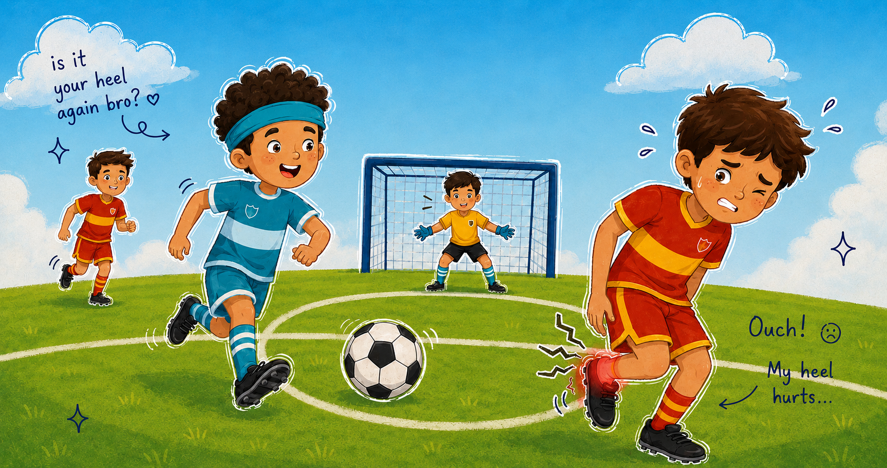
Heel pain in an active child is extremely common. In children between roughly 8 and 15 years of age, the most frequent diagnosis is calcaneal apophysitis, better known as Sever’s disease.

Despite the alarming name, Sever’s disease is not a disease in the conventional sense. It is a growth-related, load-sensitive injury affecting the developing heel.

It is particularly relevant to children who run, jump and train frequently—footballers, basketball players, runners, gymnasts, dancers, tennis players and children participating in multiple school and club sports.

In Singapore, this pattern is especially familiar because many children combine school physical education, CCAs, academies and weekend sport throughout the year. There is, however, no good published Singapore-specific prevalence figure for Sever’s disease, so claims that a particular percentage of Singaporean children develop the condition would currently be speculative.

What we do know is that Sever’s disease can substantially affect a child's ability to participate in sport, walk comfortably and enjoy normal activities. It is therefore misleading to dismiss it simply as “growing pains.” Studies examining quality of life demonstrate meaningful physical and functional effects in affected children.

The encouraging news is that Sever’s disease is self-limiting and does not normally cause permanent damage. The challenge is managing the painful period properly while the heel is still developing.


What exactly is Sever’s disease?

The heel bone is called the calcaneus.

In an adult, the posterior calcaneus is mature solid bone. In a growing child, however, the back of the calcaneus contains a developing secondary ossification centre known as the calcaneal apophysis.

An apophysis is a growth region situated where powerful muscles and tendons transmit force into bone.

The Achilles tendon attaches to the posterior calcaneus. During running and jumping, the calf muscles contract forcefully and transmit substantial loads through the Achilles tendon into this developing heel.

The calcaneal secondary ossification centre usually becomes visible at approximately seven or eight years of age and progressively matures until fusion, typically during adolescence. Radiographic research involving hundreds of children found complete fusion by approximately 15 years in the studied population, although biological maturation naturally varies between individuals.

This creates a temporary window during childhood when:

the child's athletic capacity can exceed the load tolerance of the developing heel.

That is the biological setting in which Sever’s disease occurs.


Is Sever’s really an “inflammation”?

This is where the condition becomes more scientifically interesting.

The traditional name calcaneal apophysitis implies inflammation of the calcaneal apophysis.

That explanation is probably incomplete.

Several competing but overlapping mechanisms have been proposed.

1. Traction at the Achilles attachment

The classic explanation is repetitive traction from the Achilles tendon against the developing apophysis.

During growth, bone length may increase faster than the muscle-tendon complex adapts. This may temporarily increase tension through the calf-Achilles system.

Repeated running and jumping then exposes the apophysis to thousands of loading cycles.

This mechanism remains biologically plausible and is probably important in some children.

However, it should not be interpreted as:

“Every child with Sever’s has a tight Achilles tendon.”

The evidence simply does not support that.

A 2023 systematic review examining risk and associated factors found ankle dorsiflexion restriction to be one of the most frequently investigated variables, but results across studies were inconsistent. Some children demonstrate restricted dorsiflexion; others do not.

One relatively large study of 124 affected children actually found differences in ankle motion that did not support the simplistic assumption that all affected children had shortened calf complexes.

Therefore:

Calf tightness may contribute to Sever’s disease in some children, but it should be measured rather than assumed.

2. Repetitive impact through the heel

The calcaneal apophysis does not experience Achilles traction alone.

Every time the heel hits the ground during running, landing or jumping, ground-reaction forces travel upward through the calcaneus.

This is particularly important in:

  • football
  • basketball
  • athletics
  • gymnastics
  • tennis
  • court sports
  • dance
  • repeated sprinting
  • jumping activities.

Several plantar-pressure studies have investigated whether children with Sever’s disease load their heels differently.

One case-control study involving young footballers found substantially greater heel plantar pressures and altered movement of the centre of pressure in affected children.

Other biomechanical research has been less clear. A 2019 treadmill study did not find significantly greater peak vertical ground-reaction forces or regional plantar pressures overall. Interestingly, however, children with calcaneal apophysitis ran with a slightly higher cadence and shorter step length, potentially representing an adaptation designed to reduce painful heel loading. Heel pressure during running was also associated with reported pain.

This tells us something important:

Sever’s disease is probably not caused by one abnormal force.

It is more likely a problem of cumulative mechanical load exceeding temporary tissue capacity.

3. Stress injury and bone remodelling

Perhaps the most interesting evidence comes from pathology and MRI research.

In 1995, Liberson and colleagues examined the calcaneal apophysis using radiographs, CT and histology. They identified disruption and reparative changes that supported a process of stress remodelling caused by repetitive bending forces rather than simply nonspecific inflammation.

Ogden and colleagues later examined children with persistent symptoms using MRI and proposed something even more provocative.

They observed bone-bruising-like signal changes in the developing calcaneal metaphysis and suggested that what we call Sever’s disease might in some cases behave more like a trabecular stress injury of the immature calcaneus than a pure apophysitis.

This theory remains debated.

It does, however, fit with the contemporary model:

Sever’s disease represents a spectrum of mechanically induced stress within the growing posterior calcaneus.

That spectrum may involve:

  • traction
  • compression
  • impact
  • shear
  • bending
  • bone remodelling
  • local oedema
  • irritation around the apophysis.

Calling it simply “inflammation caused by a tight Achilles” therefore probably understates the biology.


The growth plate is the weak link

Children and adults respond differently to repetitive loading.

In a skeletally mature adult, repetitive tensile loading might produce Achilles tendinopathy.

In a growing child, the tendon may be relatively strong while the developing bone-tendon interface remains vulnerable.
The weak link therefore shifts toward the apophysis.

This helps explain why several childhood conditions follow a similar pattern:

  • Sever’s disease — calcaneus
  • Osgood–Schlatter disease — tibial tuberosity
  • Sinding-Larsen–Johansson syndrome — inferior patella.

These are all examples of growing attachment sites being repeatedly loaded during sport.

Recent ultrasound research also suggests that children with Sever’s disease may demonstrate differences in Achilles tendon morphology compared with controls, further supporting the idea that the Achilles–calcaneus system should be viewed as a functional unit, although association does not establish causation.


Who gets Sever’s disease?

The typical child is approximately 8–15 years old, although the exact age depends on skeletal maturation.

It commonly appears around periods of rapid growth.

Boys have historically been reported more often than girls. A systematic review containing 1,362 reported cases found approximately 71% were boys.
However, older datasets reflect historical differences in sports participation, and Sever’s disease certainly affects girls—particularly those involved in gymnastics, dance and high-volume running and jumping sports.

Both heels may be affected.

In one classic series of 85 children, 61% had bilateral symptoms.

A more recent systematic review also demonstrated frequent bilateral presentation.


How common is it?

Exact prevalence is difficult to establish because many children are never formally diagnosed.

A general-population study from the Netherlands estimated an incidence of approximately 3.7 per 1,000 children per year.

In sports and musculoskeletal clinics, Sever’s disease has historically accounted for roughly 2–16% of paediatric musculoskeletal presentations, depending on the population studied.

A 10-year study from an elite German youth football academy confirmed that calcaneal apophysitis can produce considerable sporting time loss, with an average return-to-play time of approximately 61 days, although the enormous variation between children is important.

It therefore deserves considerably more attention than the phrase “growing pains” implies.


What increases the risk?

This is another area where medical folklore has moved faster than the science.

The best systematic review of risk factors found no single universally accepted risk factor.

The most plausible factors are the following.

Rapid growth

Growth changes the relationship between:

  • bone length
  • muscle length
  • tendon stiffness
  • body mass
  • lever arms
  • coordination
  • sports load.

A child who suddenly becomes taller and heavier may be exposing the heel to substantially more force even if the training programme itself has not changed.


Running and jumping load

High-impact sport is biologically plausible and repeatedly associated with apophyseal problems.

A large prospective study of lower-limb apophysitis published in 2025 found higher incidence among children participating in high-impact sports such as football, handball, basketball and gymnastics. Sever’s, Osgood–Schlatter and Sinding-Larsen–Johansson disease comprised virtually all cases in that cohort.

But another important nuance exists:

simply counting training sessions is not enough.

One athletic-population study unexpectedly found Sever’s disease more commonly among younger and somewhat less active children and did not identify sport type, BMI, sex or foot posture as significant risk factors in that cohort.

That is why training load must be considered alongside growth and individual tissue capacity.


Sudden increases in training

A child may tolerate three football sessions per week for months.

Problems may begin when that becomes:

  • school football
  • club football
  • weekend matches
  • athletics
  • additional conditioning.

No single session necessarily appears excessive.

The problem is cumulative loading.

This is particularly relevant when sport volume rises rapidly after:

  • school holidays
  • an off-season
  • injury
  • joining a competitive academy
  • moving into a higher age group.


Body weight

Several studies have found affected children to be heavier or to have higher BMI than population norms, and one case-control plantar-pressure study found higher BMI among affected footballers.

But other studies have found no significant relationship.

Therefore it is inappropriate to tell every child with Sever’s disease that body weight is the cause.

Mechanical loading is influenced by body mass, but BMI is one variable among many.


Flat feet and pronation

Pronation is frequently blamed for Sever’s disease.

Again, the evidence is mixed.

Some studies have found more pronated foot posture among affected children. The systematic risk-factor review identified foot alignment as a possible associated factor.

Micheli and Ireland also reported pronation as the most common associated foot condition in their original clinical series.

However, other athletic studies have failed to show foot posture as an independent risk factor.

The sensible clinical interpretation is therefore:

Pronated feet do not automatically cause Sever’s disease.

But if an individual child has substantial pronation and it appears to alter heel loading or increase symptoms, addressing that mechanical factor may be useful.

The same logic applies to unusually supinated feet.

Treatment should follow examination rather than ideology.


Tight calf muscles

This deserves repetition because it is probably the most frequently oversimplified component of Sever’s disease.

Some children genuinely have reduced ankle dorsiflexion.

For those children, calf flexibility work makes mechanical sense.

But systematic reviews show conflicting results, and affected children do not universally demonstrate equinus.

Therefore:

stretching should be prescribed when a restriction exists—not simply because the diagnosis says “Sever’s.”


What does Sever’s disease feel like?

The typical history is remarkably characteristic.

The child reports pain:

  • at the back or underside of the heel
  • during running
  • during jumping
  • after sport
  • when squeezing the heel
  • sometimes when walking after intense activity.

The child may:

  • limp
  • walk on the forefoot
  • avoid heel contact
  • stop sprinting
  • struggle with stairs
  • withdraw from sport
  • complain particularly after training rather than before it.

Symptoms usually improve with reduced loading or rest.

Pain at night while completely at rest is much less typical and deserves greater scrutiny.


How is Sever’s disease diagnosed?

For a classic presentation, Sever’s disease is primarily a clinical diagnosis.

The most useful examination manoeuvre is the calcaneal squeeze test.

The clinician compresses the posterior calcaneus from the medial and lateral sides.

Reproduction of the child's characteristic pain supports the diagnosis.

A small controlled diagnostic study reported:

  • 97% sensitivity for the squeeze test
  • 100% sensitivity for one-leg heel standing
  • 80% sensitivity for direct palpation

with 100% specificity for all three tests in that particular study population.

Those numbers should not be assumed to apply to every clinical setting because the study was small, but they reinforce the usefulness of clinical examination.

A proper assessment should also examine:

  • Achilles and calf flexibility
  • ankle range of motion
  • foot posture
  • gait
  • running mechanics where relevant
  • footwear
  • training volume
  • sporting surfaces
  • recent growth
  • strength and control of the lower limb
  • location of maximal tenderness.


Does a child need an X-ray?

Usually not simply to “prove” Sever’s disease.

This is one of the most persistent misconceptions surrounding the condition.

X-rays of healthy growing calcanei often show:

  • sclerosis
  • irregularity
  • fragmentation.

These can be normal developmental findings.

Volpon and de Carvalho Filho examined 392 healthy children and 69 boys with calcaneal apophysitis. They concluded that the sclerotic appearance of the calcaneal secondary nucleus is normal and should not be used to diagnose Sever’s disease. Fragmentation was more pronounced in symptomatic children, but it was not exclusive to them.

Similarly, the clinical diagnostic study found both increased density and fragmentation among pain-free controls.

Therefore:

An “abnormal-looking” growth plate on X-ray does not automatically mean Sever’s disease.

The diagnosis remains clinical.


When is imaging useful?

Imaging becomes important when the presentation is atypical or another diagnosis needs excluding.

This is where there is some legitimate debate.

One retrospective study of children initially thought clinically to have Sever’s disease found unexpected abnormalities on radiographs in approximately 5% of patients, including calcaneal stress fractures and bone cysts. Those authors therefore argued for routine lateral radiography.

Other reviews and clinical guidance argue that routine imaging is unnecessary in a completely typical presentation.

A sensible middle ground is:

Do not X-ray simply to look for apophyseal fragmentation.

But have a lower threshold for imaging when the clinical picture is unusual.

MRI is substantially more sensitive when concern exists for:

  • stress fracture
  • osteomyelitis
  • tumour
  • marrow pathology
  • other deep structural disease.

Ultrasound may demonstrate apophyseal and surrounding soft-tissue changes but is not ordinarily necessary for a straightforward diagnosis.


Red flags: when heel pain may not be Sever’s disease

Parents and clinicians should reconsider the diagnosis when a child has:

  • persistent pain at rest
  • significant night pain
  • fever
  • redness or marked warmth
  • unexplained swelling
  • systemic illness
  • unexplained weight loss
  • severe inability to bear weight
  • major acute trauma
  • neurological symptoms
  • progressive deterioration despite appropriate management
  • an age or skeletal maturity inconsistent with calcaneal apophysitis.

The differential diagnosis includes:

  • calcaneal stress fracture
  • Achilles tendinopathy
  • retrocalcaneal bursitis
  • plantar fascia-related pain
  • calcaneal bone cyst
  • osteoid osteoma or other tumour
  • osteomyelitis
  • juvenile inflammatory arthritis
  • tarsal coalition
  • traumatic injury.

The words “growing pains” should never be used to dismiss an atypical presentation.


So what actually works?

This is the most important part of the discussion.

A 2024 systematic review examining randomized controlled trials concluded that conservative interventions—including orthotic devices, heel cups/lifts, exercise therapy and taping—can improve symptoms, but the studies are heterogeneous and no single treatment has emerged as universally superior.

Earlier systematic reviews reached a similar conclusion: there is evidence for several interventions, but considerably less high-quality evidence than the confidence with which treatments are sometimes recommended.

The best treatment therefore targets the child's load problem and individual mechanical contributors.

1. Load management

Evidence verdict: ESSENTIAL

This does not necessarily mean complete rest.

The objective is to bring mechanical exposure temporarily below the painful heel's current tolerance.

That may involve reducing:

  • sprinting
  • repeated jumping
  • high-volume running
  • double training sessions
  • hard-surface activity.

A child whose heel hurts after two hours of football may tolerate:

  • 45 minutes
  • technical drills
  • swimming
  • cycling
  • upper-body conditioning.

In other words:

Modify the load rather than automatically removing the child from all physical activity.

This is particularly relevant because some studies have successfully treated children while they continued participating in sport.

If the child is visibly limping or changing running technique significantly because of pain, however, the load is clearly too high.

2. Heel cups

Evidence verdict: GOOD SHORT-TERM OPTION

Heel cups have some of the most mechanistically convincing evidence.

Perhamre and colleagues demonstrated that a rigid heel cup could:

  • support the heel fat pad
  • increase functional heel-pad thickness
  • reduce heel peak pressure
  • reduce pain.

In a randomized crossover study involving 51 boys, the heel cup provided markedly better sporting pain relief than a heel wedge and was preferred by more than 75% of participants.

This is particularly interesting because the benefit may not simply result from “relaxing the Achilles.”

The cup mechanically supports the heel and changes impact attenuation.

That fits the broader load-based model of Sever’s disease.

3. Heel lifts

Evidence verdict: CAN HELP, PARTICULARLY SHORT TERM

Heel lifts slightly plantarflex the ankle inside the shoe and may reduce tensile demand through the Achilles-apophyseal complex.

They can also alter heel loading.

A randomized trial comparing:

  • wait-and-see
  • heel raise
  • supervised physiotherapy

found all three approaches improved symptoms.

At six weeks, children using heel raises showed some functional and satisfaction advantages.

By three months, however, there were no clinically important differences between the groups.

Another large randomized trial followed 124 children and found a modest short-term physical-function advantage for heel raises compared with prefabricated orthoses at one and two months. That advantage disappeared by six and twelve months.

Heel lifts therefore appear useful primarily as a temporary symptom-modifying tool, rather than a cure.

4. Foot orthoses

Evidence verdict: USEFUL IN SELECTED CHILDREN; STRONGER EVIDENCE FOR CUSTOM DEVICES THAN MANY PEOPLE REALISE

This area requires careful interpretation because the studies used very different devices.

A 2021 randomized trial involving 208 children compared:

  • custom polypropylene foot orthoses
  • off-the-shelf heel lifts.

Both groups improved.

But the custom-orthosis group improved substantially more.

Pain on the visual analogue scale fell by approximately 68.6% in the custom-orthosis group, with substantially greater improvement in pressure-pain thresholds than in the heel-lift group.

That is an important result.

It does not, however, mean that every child with Sever’s disease requires custom orthotics.

Why?

Because orthoses become most logical when the examination identifies a mechanical reason for using them—for example:

  • significant pronation
  • unusual rearfoot mechanics
  • excessive heel loading
  • asymmetry
  • symptoms strongly influenced by foot function.

A separate 2016 randomized trial did not show prefabricated orthoses to be superior to simple heel raises.

The key distinction is therefore:

“Orthotic” is not one treatment.

A generic prefabricated insert and an individually prescribed custom device designed around a child's biomechanics are fundamentally different interventions.

5. Shoes

Evidence verdict: IMPORTANT FOR COMFORT AND LOAD MANAGEMENT, BUT BUYING A NEW SHOE IS NOT A CURE

Shoes influence:

  • heel cushioning
  • heel-to-toe drop
  • stability
  • impact attenuation
  • how an orthotic or heel cup functions.

Children with acute Sever’s symptoms often tolerate a cushioned sports shoe better than:

  • barefoot activity
  • very flat shoes
  • hard school shoes
  • thin minimalist shoes
  • worn-out football boots.

But interestingly, the 12-month randomized footwear trial did not find that prescribed new athletic footwear produced a major independent long-term advantage.

So the message is not:

Buy an expensive shoe and Sever’s disease disappears.

The better message is:

Avoid footwear that repeatedly aggravates the heel, and create a comfortable mechanical environment while the apophysis settles.

What about football boots?

Football deserves particular attention because it appears repeatedly in Sever’s literature.

Studs provide considerably less cushioning than most running shoes, and football combines:

  • sprinting
  • acceleration
  • deceleration
  • jumping
  • repeated forefoot loading
  • long training duration.

A child may therefore report:

“My heel is fine in school shoes but hurts badly after football.”

That pattern is entirely plausible.

A temporary heel cup or appropriate insert, where it fits safely within the boot, may make a significant difference.

Training volume must still be considered.

6. Stretching

Evidence verdict: USE WHEN INDICATED—NOT AUTOMATICALLY

Stretching is probably the most universally prescribed treatment for Sever’s disease.

Yet surprisingly, high-quality evidence specifically proving that stretching itself cures Sever’s disease is limited.

This is important.

If examination demonstrates genuine gastrocnemius or soleus restriction, restoring appropriate dorsiflexion is reasonable.

But aggressively stretching every child merely because the diagnosis is Sever’s disease has weak scientific justification.

Overly aggressive calf stretching may also temporarily increase tensile loading through an already sensitive posterior heel.

Therefore:

Stretch a restriction—not a diagnosis.

7. Strengthening and physiotherapy

Evidence verdict: USEFUL, PARTICULARLY WHEN FUNCTIONAL DEFICITS EXIST

Physiotherapy may include:

  • calf strengthening
  • progressive heel raises
  • ankle control
  • balance
  • hip and lower-limb strength
  • gradual return to running
  • mobility where restricted
  • load education.

The randomized trial comparing physical therapy with heel raises and observation found all strategies effective by three months. Physiotherapy produced some earlier functional advantages according to parent-reported outcomes but was not ultimately superior for pain.

That suggests physiotherapy should not be viewed as a magical treatment for the growth plate itself.

Its value lies in restoring the capacity of the entire kinetic chain to tolerate sport.


Should children perform eccentric Achilles exercises?

Adult Achilles tendinopathy protocols are sometimes transferred directly to children with Sever’s disease.

That is questionable.

Adult Achilles tendinopathy and paediatric calcaneal apophyseal pain are different biological conditions.

Eccentric exercise formed part of the physical-therapy intervention in one randomized Sever’s trial, but it was not clearly superior to heel lifts or observation at three months.

Exercise should therefore be progressive and age-appropriate rather than copied blindly from adult tendinopathy programmes.

8. Kinesio taping

Evidence verdict: POSSIBLE ADJUNCT; NOT A PRIMARY TREATMENT

A randomized study comparing genuine Kinesio tape with sham taping found both groups experienced substantial pain improvement.

Pain scores were not significantly different between groups, although some functional scores favoured Kinesio taping at selected follow-ups.

That means taping may help certain children feel or function better, but claims that tape is treating the underlying growth plate are unsupported.

Use it as an adjunct if helpful—not as the foundation of management.

9. Ice

Evidence verdict: MAY HELP PAIN; DOES NOT FIX THE CAUSE

Ice is inexpensive and can reduce discomfort following sport.

But there is no convincing evidence that icing changes the natural biological course of Sever’s disease.

If a child likes it and obtains relief, it is reasonable.

If they dislike it, treatment does not fail because ice was omitted.

10. Anti-inflammatory medication

Evidence verdict: SHORT-TERM PAIN RELIEF ONLY

Ibuprofen and other anti-inflammatory medication are frequently recommended.

They may reduce pain.

However, Sever’s disease is fundamentally a mechanical load problem in developing tissue, not simply an inflammatory disease.

Medication should therefore never be used to allow a child to repeatedly train through significant pain.

Medication in children should also be used according to appropriate paediatric medical advice, dosing and contraindications.

11. Immobilisation or a walking boot

Evidence verdict: RESERVED FOR SEVERE CASES

Most children do not require immobilisation.

A boot may occasionally be appropriate when:

  • walking is painful
  • the child is significantly limping
  • symptoms are severe
  • ordinary load reduction has failed
  • a stress injury is suspected.

The purpose is temporary unloading.

Routine prolonged immobilisation for ordinary Sever’s disease is unnecessary and can lead to avoidable weakness and deconditioning.

12. Shockwave therapy

Evidence verdict: EXPERIMENTAL FOR SEVER’S DISEASE

Extracorporeal shockwave therapy has established applications in several adult musculoskeletal disorders.

Evidence in growing children with apophyseal injuries is extremely limited.

A retrospective case series included only seven children with Sever’s disease among 22 young athletes treated for different apophyseal conditions.
 
Results were encouraging, but a seven-patient uncontrolled subgroup cannot establish effectiveness or long-term safety.

Therefore ESWT should not currently be presented to parents as an established first-line Sever’s treatment.

Much stronger evidence is required.


What does NOT appear justified?

Based on current evidence, several common approaches deserve caution.

Complete sports cessation for every child

Not always necessary.

Some children can remain active if load is reduced sufficiently and pain is controlled.

Several heel-cup studies allowed children to maintain high sporting activity while symptoms improved.

Treating an X-ray instead of the child

Fragmentation and sclerosis of the calcaneal apophysis can occur normally.

They should not dictate treatment in isolation.

Telling every child their calf is too tight

Not evidence-based.

Measure ankle mobility first.

Assuming flat feet caused the problem

Pronation may be relevant in an individual child but is not a universal cause.

Medication without changing load

Pain medication can mask symptoms while the mechanical overload continues.

It does not address the primary problem.

Aggressive stretching through heel pain

Stretching should not repeatedly reproduce significant apophyseal pain.

Promising that one treatment “cures” Sever’s

No intervention has demonstrated universal superiority.

The biology resolves as the apophysis matures.

Treatment primarily helps the child control symptoms and maintain function during that period.


A practical evidence-based treatment framework

For most children, treatment can be organised into four stages.

Stage 1 — Reduce the mechanical irritation

For one to several weeks:

  • temporarily reduce the most painful running and jumping volume
  • avoid training through a limp
  • use comfortable cushioned footwear
  • consider a heel cup or heel lift
  • use ice for comfort if helpful.

The goal is not necessarily complete inactivity.

The goal is pain control without repeatedly exceeding tissue tolerance.


Stage 2 — Identify why this particular heel is overloaded

Assess:

  • ankle dorsiflexion
  • calf flexibility
  • foot posture
  • pronation/supination
  • heel pressure
  • gait
  • running technique
  • strength
  • recent growth
  • body mass changes
  • footwear
  • weekly sporting load.

This is where individualised treatment begins.


Stage 3 — Restore capacity

Where appropriate:

  • calf flexibility
  • progressive calf strengthening
  • intrinsic foot strength
  • balance
  • lower-limb strength
  • running progression.

If substantial foot mechanics contribute to loading, consider an appropriately selected orthotic intervention.


Stage 4 — Gradually restore sports load

Return should be based primarily on function rather than an arbitrary number of weeks.

The child should progressively demonstrate:

  • normal walking
  • no significant limp
  • comfortable hopping
  • comfortable running
  • comfortable acceleration
  • comfortable sport-specific drills.

A small amount of transient discomfort may sometimes be acceptable during rehabilitation, but there is currently no universally validated Sever’s-specific pain-monitoring threshold.

A useful principle is:

symptoms should settle quickly and should not be progressively worse later that day or the following morning.

A 2026 feasibility study is now specifically investigating exercise therapy combined with pain-guided activity modification, which reflects the direction modern management is moving.


How long does Sever’s disease take to recover?

There is no single answer.

Mild cases may improve within several weeks.

Others fluctuate for several months.

The German elite-football study found an average return-to-play time of approximately 61 days, but with very large variation between players. Recurrent cases took longer.

The older Micheli series reported return to sport at approximately two months with conservative management.

Importantly, symptoms can recur.

Why?

Because treatment does not instantly mature the growth plate.

A child may improve, increase sport dramatically and overload the same biologically vulnerable structure again.

Recurrence therefore does not necessarily mean something has gone wrong.

It often means:

Load once again exceeded capacity.


Does Sever’s disease permanently damage the heel?

In ordinary cases, no.

The condition is considered self-limiting and usually disappears once the calcaneal apophysis matures and fuses.

There is no convincing evidence that routine Sever’s disease produces chronic adult heel pathology.

That is reassuring.

But “it eventually goes away” should not be used as an excuse to ignore pain for months.

Repeated pain can interfere with:

  • sport
  • movement
  • confidence
  • school activities
  • physical fitness
  • quality of life.

Good management therefore matters even when the long-term prognosis is excellent.


Can Sever’s disease be prevented?

There is no proven prevention programme.

But several principles are sensible.

Avoid sudden training spikes
A child's load should increase progressively.

Watch periods of rapid growth
A programme tolerated six months earlier may suddenly become too demanding.

Use appropriate footwear for the activity
Especially during high-volume running and jumping.

Address genuine mobility restrictions
Not every child requires stretching, but restricted ankle function should not be ignored.

Build lower-limb strength
The stronger and better coordinated the kinetic chain, the better it can distribute sporting load.

Do not normalise persistent pain
A child repeatedly limping after sport deserves assessment.


The most important concept: load versus capacity

The best way to understand Sever’s disease is with a simple equation:

Mechanical load > temporary tissue capacity = pain

A growing calcaneus has a temporary biological limit.

Running, jumping and Achilles traction supply the load.

Rapid growth, body mass, foot mechanics, footwear, strength and recovery modify the relationship.

Treatment therefore attempts to achieve two things:

Reduce excessive load
and
Improve the child's ability to tolerate load.

Heel cups, orthoses, footwear and training modification primarily change the load side of the equation.

Strength, mobility and progressive rehabilitation influence the capacity side.

Growth eventually changes the tissue itself.

This is a far more useful model than telling a child:

“Your Achilles is pulling on your growth plate. Stop sport and stretch.”


What parents should remember

Sever’s disease is common.

It is painful but usually temporary.

It is not simply a mysterious “growing pain.”

It appears to be a genuine mechanical stress response within the developing heel.

The Achilles tendon matters—but so do impact forces, growth, sport volume and individual biomechanics.

X-rays are not routinely required to confirm the diagnosis, and fragmentation of the apophysis is not diagnostic by itself.

Treatment should be individualised.

For some children, load reduction and a heel cup may be enough.

Others benefit from a heel lift, rehabilitation or an orthotic intervention.

Children with marked biomechanical abnormalities may require more detailed mechanical management.

Most importantly:

Persistent heel pain should be managed rather than ignored, but children do not automatically need to abandon sport completely.
The objective is to keep the growing athlete active while respecting the biological limits of the developing heel.


Evidence summary: what works?


Load modification | Strong clinical rationale | Foundation of treatment

Heel cups | Moderate evidence | Good option for reducing heel pressure and sporting pain

Heel lifts | Moderate evidence | Useful short-term symptom modifier

Custom orthoses | Moderate evidence; one large positive RCT | Particularly reasonable when individual biomechanics justify them

Prefabricated orthoses | Mixed evidence | Not clearly superior to heel lifts

Supportive/cushioned footwear | Reasonable adjunct | Helpful for comfort; new shoes alone are not a cure

Physiotherapy | Moderate evidence | Useful where strength/mobility deficits exist

Calf stretching | Conditional | Appropriate if ankle/calf restriction is actually present

Kinesio taping | Low–moderate evidence | Possible adjunct; limited effect beyond placebo for pain

Ice | Symptomatic only | Fine for comfort

NSAIDs | Symptomatic only | Short-term pain control; not a mechanical solution

Immobilisation | Low-level evidence | Reserve for severe painful cases

ESWT | Insufficient evidence | Experimental in paediatric apophysitis

Surgery | No role in routine Sever’s disease | Condition is self-limiting


Book An Appointment

Disclaimer: The word "treatment" in this article refers to the care and management of a patient’s health to prevent, cure, or improve a condition. Treatment results vary and do not necessarily indicate a cure. This article is for informational and educational purposes only and does not constitute medical advice.

About the Author

Mark B. Reyneker, BTech (Podiatry), MSc (Palaeontology) is a podiatrist and Founder & Clinical Director of Family Podiatry Centre, with more than 25 years of clinical experience across South Africa, Malaysia and Singapore. His clinical interests include foot and lower-limb pain, gait and biomechanics, sports-related foot conditions, orthotic therapy and footwear.

Alongside his clinical practice, Mark conducts research into human gait and foot biomechanics. His MSc research at the University of the Witwatersrand investigated human propulsion and the structural properties of the metatarsals. He is also the inventor of A Foot Orthotic, an orthotic technology developed through an international patent family.


References and key peer-reviewed literature

1. James AM, Williams CM & Haines TP (2013). Effectiveness of interventions in reducing pain and maintaining physical activity in children and adolescents with calcaneal apophysitis (Sever’s disease): a systematic review. Journal of Foot and Ankle Research, 6, 16. Article / PubMed record

2. Nieto-Gil P et al. (2023). Risk factors and associated factors for calcaneal apophysitis (Sever’s disease): a systematic review. BMJ Open, 13, e064903. Article / PubMed record

3. Hernández-Lázaro P et al. (2024). Conservative Treatment of Sever’s Disease: A Systematic Review. Journal of Clinical Medicine, 13, 1391. Full article

4. Fares MY et al. (2023). Clinical and Diagnostic Characteristics of Calcaneal Apophysitis: A Systematic Review and Thematic Analysis. Journal of the American Podiatric Medical Association, 113. Article / PubMed record

5. Scharfbillig RW, Jones S & Scutter SD (2008). Sever’s disease: what does the literature really tell us? Journal of the American Podiatric Medical Association, 98, 212–223. Article / PubMed record

6. Hendrix CL (2005). Calcaneal apophysitis (Sever disease). Clinics in Podiatric Medicine and Surgery, 22, 55–62. Article / PubMed record

7. Micheli LJ & Ireland ML (1987). Prevention and management of calcaneal apophysitis in children: an overuse syndrome. Journal of Pediatric Orthopaedics, 7, 34–38. Article / PubMed record

8. Liberson A et al. (1995). Remodeling of the calcaneus apophysis in the growing child. Journal of Pediatric Orthopaedics B, 4, 74–79. Article / PubMed record

9. Ogden JA et al. (2004). Sever’s injury: a stress fracture of the immature calcaneal metaphysis. Journal of Pediatric Orthopaedics, 24, 488–492. Article / PubMed record

10. Volpon JB & de Carvalho Filho G (2002). Calcaneal apophysitis: a quantitative radiographic evaluation of the secondary ossification center. Archives of Orthopaedic and Trauma Surgery, 122, 338–341. Article / PubMed record

11. Hoşgören B, Köktener A & Dilmen G (2005). Ultrasonography of the calcaneus in Sever’s disease. Indian Pediatrics, 42, 801–803. Article / PubMed record

12. Perhamre S et al. (2013). Sever’s injury: a clinical diagnosis. Journal of the American Podiatric Medical Association. Article / PubMed record

13. Rachel JN et al. (2011). Is radiographic evaluation necessary in children with a clinical diagnosis of calcaneal apophysitis (Sever disease)? Journal of Pediatric Orthopaedics. Article / PubMed record

14. Becerro de Bengoa Vallejo R et al. (2011). Plantar pressures in children with and without Sever’s disease. Journal of the American Podiatric Medical Association, 101, 17–24. Article / PubMed record

15. López-López D et al. (2018). Slow velocity of the center of pressure and high heel pressures may increase the risk of Sever’s disease: a case-control study. BMC Pediatrics. Full article

16. McSweeney S, Reed LF & Wearing SC (2019). Vertical ground reaction forces during gait in children with and without calcaneal apophysitis. Gait & Posture, 71, 126–130. Article / PubMed record

17. Scharfbillig RW, Jones S & Scutter S (2011). Sever’s disease: a prospective study of risk factors. Journal of the American Podiatric Medical Association, 101, 133–145. Article / PubMed record

18. James AM et al. (2015). Factors Associated with Pain Severity in Children with Calcaneal Apophysitis (Sever Disease). Journal of Pediatrics, 167, 455–459. Article / PubMed record

19. Wiegerinck JI et al. (2014). Incidence of calcaneal apophysitis in the general population. European Journal of Pediatrics, 173, 677–679. Article / PubMed record

20. Perhamre S et al. (2011). Sever’s injury: treatment with insoles provides effective pain relief. Scandinavian Journal of Medicine & Science in Sports, 21, 819–823. Article / PubMed record

21. Perhamre S et al. (2011). Sever’s injury; treat it with a heel cup: a randomized, crossover study with two insole alternatives. Scandinavian Journal of Medicine & Science in Sports, 21, e42–e47. Article / PubMed record

22. Perhamre S et al. (2012). A heel cup improves the function of the heel pad in Sever’s injury: effects on heel pad thickness, peak pressure and pain. Scandinavian Journal of Medicine & Science in Sports, 22, 516–522. Article / PubMed record

23. Wiegerinck JI et al. (2016). Treatment of Calcaneal Apophysitis: Wait and See Versus Orthotic Device Versus Physical Therapy: A Pragmatic Therapeutic Randomized Clinical Trial. Journal of Pediatric Orthopaedics, 36, 152–157. Article / PubMed record

24. James AM, Williams CM & Haines TP (2016). Effectiveness of footwear and foot orthoses for calcaneal apophysitis: a 12-month factorial randomised trial. British Journal of Sports Medicine, 50, 1268–1275. Article / PubMed record

25. James AM, Williams CM & Haines TP (2016). Health related quality of life of children with calcaneal apophysitis: child & parent perceptions. Health and Quality of Life Outcomes, 14, 95. Article / PubMed record

26. Kuyucu E et al. (2017). Assessment of the kinesiotherapy’s efficacy in male athletes with calcaneal apophysitis. Journal of Orthopaedic Surgery and Research. Article / PubMed record

27. Alfaro-Santafé J et al. (2021). Effectiveness of Custom-Made Foot Orthoses vs. Heel-Lifts in Children with Calcaneal Apophysitis (Sever’s Disease): A CONSORT-Compliant Randomized Trial. Children, 8, 963. Article / PubMed record

28. Feyzioğlu Ö, Öztürk Ö & Muğrabi S (2021). Effects of custom-made insoles on foot pressure redistribution, gait parameters, and pain in calcaneal apophysitis. Prosthetics and Orthotics International, 45, 532–537. Article / PubMed record

29. Scharfbillig et al./German youth football academy cohort (2022). Incidence of calcaneal apophysitis and return-to-play in adolescent athletes of a German youth soccer academy: a retrospective study of 10 years. Article / PubMed record

30. Sweeney EA et al. (2023). Comparison of Braces for Treatment of Sever’s Disease in Barefoot Athletes: A Randomized Clinical Trial. Journal of Athletic Training, 58, 437–444. Article / PubMed record

31. Hanlon SL, Whitney KE & DeJong Lempke AF (2024). Youth Athletes With Sever’s Disease Exhibit Altered Achilles Tendon Ultrasound Characteristics: A Retrospective Case-Control Study. Journal of Ultrasound in Medicine, 43, 1303–1312. Article / PubMed record

32. Wedderkopp N et al. (2026). Incidence of and Risk Factors for Lower Extremity Apophysitis in Children and Adolescents. Sports Medicine, 56, 793–803. Full article

33. Hanlon SL et al. (2026). The feasibility of a novel exercise therapy and activity modification intervention for patients with Sever’s disease. Article / PubMed record

Frequently Asked Questions

Sever’s disease (calcaneal apophysitis) is a common, growth-related heel condition in active children, typically between 8 and 15 years old. It occurs when repetitive running, jumping, and physical activity overload the developing growth plate at the back of the heel bone (calcaneus).

No. Despite the word "disease" in its name, Sever’s disease is not an infection or a illness, nor is it contagious. It is an overuse injury related to growth and sport-related stress on the immature heel bone.

It is caused by cumulative mechanical stress—such as repetitive impact from running and jumping or traction from the Achilles tendon pulling against the calcaneal growth plate. Rapid growth spurts can make the tissue around the heel temporarily more vulnerable to these forces.

Symptoms include heel pain during or after physical activity, tenderness when squeezing the sides of the heel, limping after sports, walking on toes to avoid putting weight on the heel, and localized stiffness or soreness after waking up or sitting for long periods.

A qualified podiatrist or healthcare professional usually diagnoses Sever’s disease through a thorough clinical and physical examination, including the "heel squeeze test." X-rays are generally not required unless the podiatrist needs to rule out fractures, bone cysts, or other conditions.

No. While it occurs during growth, treating it merely as normal "growing pains" can lead to unnecessary discomfort, decreased sports performance, and prolonged inactivity. Proper load management and treatment help active children manage the symptoms comfortably.

Yes. It is very common for Sever’s disease to present bilaterally (in both heels), occurring in over 60% of cases due to equal sports participation and load distribution across both lower limbs.

High-impact, running, and jumping sports are most frequently associated with Sever’s disease. These include football, basketball, gymnastics, athletics, tennis, dance, and track and field.

The condition is self-limiting and resolves when the growth plate in the heel fuses (typically around age 14 to 15). However, individual painful episodes usually last anywhere from a few weeks to several months, depending on how effectively activity levels and heel loads are managed.

No. Sever’s disease typically leaves no permanent damage or long-term disability. Once the growth plate fully matures and closes, symptoms disappear completely.

Rarely. Total rest is usually unnecessary and counterproductive. Instead, podiatrists recommend temporary "load management"—modifying training volume, frequency, or intensity while providing symptomatic relief so the child can safely stay active.

Evidence-based management focuses on: Temporary activity adjustment (load modification) Cushioning, padded heel cups, or elevated heel lifts Podiatric custom orthotics to optimize foot mechanics and reduce heel strain Targeted calf and Achilles stretches or strengthening exercises (when appropriate) Ice application post-activity for temporary pain control

Yes. Padded heel cups elevate the heel slightly to reduce tension from the Achilles tendon and absorb ground impact forces. Custom orthotics can help distribute foot pressures evenly across the foot, particularly if the child has underlying biomechanical issues like flat feet or excessive pronation.

Not always immediately. While tight calves can contribute to traction on the heel, stretching an intensely inflamed growth plate can sometimes aggravate the pain. Calf stretching should be introduced carefully and progressively guided by a podiatrist.

Children should wear supportive, well-cushioned shoes with a mild heel-to-toe drop (rather than completely flat footwear). Avoid barefoot walking on hard surfaces, minimalist shoes, or worn-out sports cleats during symptomatic flare-ups.

Football or rugby cleats typically have minimal heel cushioning and thin soles, which increase ground impact directly onto the heel. Additionally, the stud placement can alter foot mechanics and increase Achilles tension.

Sever’s disease: Pain is located directly at the back/sides of the heel bone in children aged 8–15. Achilles tendinopathy: Pain is located higher up in the Achilles tendon body rather than the bone. Plantar fasciitis: Pain is underneath the heel arch and is far more common in adults than growing children.

Applying ice packs wrapped in a towel for 10–15 minutes after activity can reduce localized discomfort. Over-the-counter pain medications may offer temporary relief during severe flare-ups, but they should only be used under medical guidance and do not replace proper load management.

Yes. Symptoms can resurface during subsequent growth spurts or when sports training volume sharply increases (e.g., starting a new sports season or intensive training camp). Continuing load management and supportive footwear helps prevent recurrences.

You should see a podiatrist if your child limps, complains of persistent heel pain during or after physical activity, shows night pain, or if there is visible swelling, redness, or severe pain that prevents normal weight-bearing.
All rights reserved | Blog