Published: 17 August 2026 | Last reviewed: 16 September 2026
A plantar wart is not simply a lump of hard skin that needs to be cut away. It is a localised viral infection involving the living cells of the epidermis—the outer layer of the skin.
The infection is caused by certain types of human papillomavirus, better known as HPV. Once HPV enters the skin, it alters the normal behaviour of skin cells so that they multiply, produce excess keratin and form the thickened lesion recognised as a wart.
Plantar warts can be particularly difficult to treat because the skin on the sole is thick, the lesion is repeatedly compressed during walking, and the virus has evolved several effective ways of avoiding detection by the immune system. Treatment may therefore require more than simply destroying the visible surface.
There is no single treatment that is fast, painless, suitable for every patient and guaranteed to prevent recurrence. Choosing the most appropriate option requires consideration of the patient’s age, pain tolerance, general health, number of warts, location of the lesion and previous treatment history. (Zhu et al., 2022; Sterling et al., 2014).
What is a plantar wart?
A plantar wart—also called a verruca plantaris or simply a verruca—is a benign growth caused by HPV infection on the sole of the foot.
The word plantar refers to the bottom of the foot. Plantar warts are frequently found beneath the heel, forefoot, toes or other areas exposed to pressure.
The HPV types most commonly associated with plantar warts include HPV-1, HPV-2, HPV-4, HPV-27 and HPV-57. These are generally considered cutaneous, low-risk HPV types. They are different from the high-risk genital HPV types associated with cervical and other cancers. Ordinary plantar warts are therefore not normally considered precancerous lesions. (Zhu et al., 2022).
Two broad clinical patterns are commonly seen:
- Solitary plantar wart
This is a single, relatively well-defined lesion. It may develop a thick covering of callus and become painful when standing or walking.
- Mosaic plantar warts
These consist of numerous smaller warts that merge into a broader plaque. Mosaic warts can be difficult to eradicate because a relatively large area of skin is infected. Treating the entire area aggressively may also create an unnecessarily large wound.
Plantar warts do not have roots
Patients sometimes describe the dark dots inside a wart as “roots”. This is understandable, but medically inaccurate.
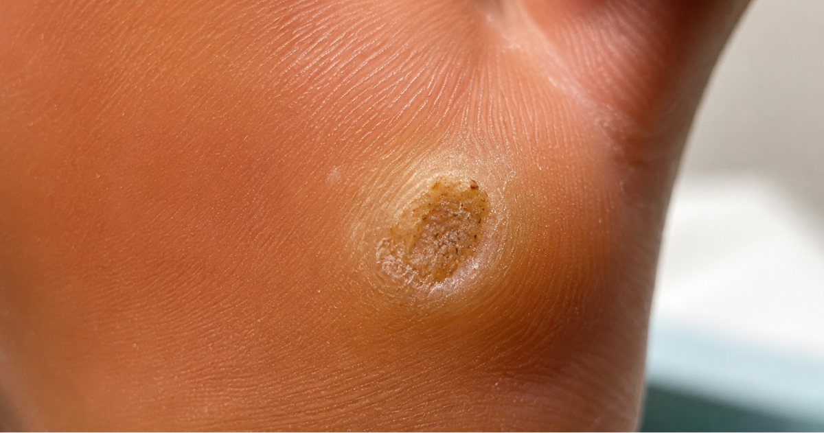
A plantar wart does not grow a root into the muscle, tendon or bone. The dark red, brown or black dots are usually small blood vessels that have become dilated, compressed or thrombosed within the wart. The lesion remains within the epidermis, although thickened keratin and pressure from standing can make it appear to extend deeply into the foot.
This distinction matters because digging for a supposed root can cause bleeding, infection and scarring without necessarily eliminating all HPV-infected cells.
Book An AppointmentWhat type of virus causes plantar warts?
HPV belongs to the Papillomaviridae family. It is a small, non-enveloped virus containing circular, double-stranded DNA.
The HPV genome contains only a limited number of genes. Rather than carrying all the machinery needed to reproduce independently, HPV takes control of a person’s keratinocytes—the cells that make up most of the epidermis—and uses the cellular machinery inside them.
HPV is highly adapted to stratified squamous epithelium. This is the type of layered tissue found in the skin and in certain mucous membranes.
The viral life cycle is closely linked to the normal maturation of a keratinocyte. As the infected skin cell moves from the deeper epidermis towards the surface, different HPV genes are switched on at different stages.
This differentiation-dependent life cycle is one reason HPV is difficult to reproduce in conventional laboratory systems and difficult to attack with a single medication. (Doorbar et al., 2015; Graham, 2017).
How does HPV enter the foot?
HPV cannot normally pass efficiently through completely intact skin. Infection generally begins when viral particles reach the deeper epidermis through a tiny area of damage.
Possible entry points include:
- Microscopic cracks caused by dry skin
- Friction from footwear
- Minor abrasions
- Scratches or picked skin
- Macerated skin that has become soft from prolonged moisture
- Trauma around an existing callus or blister
The virus infects cells close to the basal layer of the epidermis. These are long-lived cells capable of dividing and producing new generations of keratinocytes.
A person may acquire HPV weeks or months before a visible wart develops. The exact incubation period varies, and it is usually impossible to determine precisely when or where an individual became infected. Exposure does not always produce a wart. Whether a lesion develops depends on the amount of virus, the condition of the skin barrier, the HPV genotype and the effectiveness of the individual’s immune response.
What happens inside the skin?
Stage 1: Infection of a basal keratinocyte
After entering through a microscopic break, HPV reaches a basal keratinocyte. The viral DNA enters the cell nucleus and generally remains as a separate circular piece of DNA called an episome.
In an ordinary benign cutaneous wart, the viral DNA does not usually need to integrate into the human chromosomes.
Stage 2: Maintenance of the viral genome
The infected basal cell divides. HPV must preserve copies of its DNA as the cell produces daughter cells.
Early viral proteins, including E1 and E2, help regulate viral DNA replication. Other early proteins alter cell signalling and encourage cells that would normally slow down or stop dividing to continue through the cell cycle.
HPV E6 and E7 proteins are well known for their effects on cell-cycle regulation. The E6 and E7 proteins produced by wart-associated cutaneous HPV types generally have much weaker cancer-producing activity than those of high-risk mucosal HPV types.
Stage 3: Abnormal keratinocyte proliferation
As infected cells divide and move upwards through the epidermis, HPV disrupts their normal maturation. This produces several characteristic changes:
- Thickening of the epidermis
- Excess keratin production
- Elongation and distortion of the epidermal projections
- Increased prominence of small capillaries
- Interruption of the normal skin lines
The visible wart is therefore made primarily from the patient’s own altered keratinocytes rather than being a solid collection of virus.
Stage 4: Production of new viral particles
HPV delays large-scale production of its structural proteins until infected keratinocytes have moved into the upper layers of the epidermis.
The late viral proteins L1 and L2 form the outer shell, or capsid, of newly assembled viral particles. Mature viral particles are released as superficial keratinocytes naturally shed from the skin.
This is a quiet process. HPV does not usually burst the infected cells open, enter the bloodstream or produce the intense inflammation associated with many acute viral infections. This allows the virus to reproduce while attracting relatively little immune attention. (Zhou et al., 2019; Mlynarczyk-Bonikowska et al., 2024).
Why do plantar warts grow inwards?
A wart does not truly choose to grow inward. It is compressed inward by body weight, footwear and ground-reaction forces. On a finger, a wart can project outwards and form a raised, cauliflower-like surface.
On the sole, repeated pressure flattens the lesion and pushes the thickened epidermis towards the more sensitive tissues beneath it. A surrounding callus may then develop in response to pressure. This combination of viral tissue, hard keratin and mechanical compression can make the lesion painful.
The wart may feel as though a small stone has become embedded in the foot even though the infection remains primarily epidermal.
Why can a plantar wart hurt?
Pain is not caused directly by the virus attacking a nerve. It usually results from:
- Thickened tissue being driven into the foot during weight-bearing
- Increased pressure around the wart
- Distortion of sensitive tissues beneath the epidermis
- Small areas of bleeding or thrombosis
- Fissuring within a heavily callused lesion
- Secondary irritation after attempted treatment
Pain may be especially noticeable when a lesion sits beneath the heel, a metatarsal head or the tip of a toe. A traditional clinical observation is that warts may hurt more when squeezed from side to side, whereas corns may hurt more with direct downward pressure. This can be helpful, but it is not sufficiently reliable to make the diagnosis on its own.
How HPV hides from the immune system
The persistence of a wart is not necessarily evidence that the patient has a generally “weak” immune system. HPV is unusually good at remaining inconspicuous. Several viral behaviours contribute to immune evasion.
The infection remains in the epidermis
The epidermis has no direct blood supply. HPV does not normally spread through the bloodstream during a cutaneous wart infection.
This limits the exposure of viral material to circulating immune cells.
HPV does not usually destroy infected cells by lysis
Many viruses reproduce until the infected cell ruptures. Cell rupture releases inflammatory signals that attract the immune system.
HPV instead coordinates its replication with the normal maturation and shedding of keratinocytes.
New viral particles leave with superficial skin cells, creating much less alarm.
Viral proteins are produced strategically
The most immunogenic structural proteins are mainly produced in superficial epidermal layers rather than in the basal cells where long-term infection is maintained.
The virus can therefore maintain its genome in deeper cells while limiting the amount of viral antigen presented to immune surveillance.
HPV interferes with antiviral signalling
HPV proteins can suppress interferon pathways, antigen processing, inflammatory signalling and communication between infected keratinocytes and immune cells.
Different HPV types use somewhat different mechanisms, but the overall effect is delayed immune recognition and the possibility of persistent infection. (Bordignon et al., 2017; Zhou et al., 2019).
Why do some plantar warts disappear without treatment?
A wart may persist for months and then suddenly flatten and disappear. This usually means that the immune system has begun to recognise viral antigens and has mounted an effective cell-mediated response against infected keratinocytes.
Spontaneous resolution is particularly common in children. Approximately two-thirds of childhood warts may eventually disappear without destructive treatment, although the time required can be one or two years and occasionally longer. Waiting may not be practical when a wart is painful, spreading, interfering with sport or causing distress. (Soenjoyo et al., 2016; Sterling et al., 2014).
The HPV genotype may also influence the likelihood of spontaneous clearance and the response to treatment. Research has found that plantar warts associated with different HPV types do not all behave identically. (Bruggink et al., 2013).
Why can plantar warts return after treatment?
Recurrence does not necessarily mean that a wart had an undiscovered root. Possible reasons include:
- Residual infected keratinocytes: The visible lesion may have been removed while microscopic infected cells remained at its edge or base
- Subclinical infection: HPV may be present in adjacent skin before another lesion becomes visible
- Incomplete immune clearance: A destructive treatment removes tissue but may not produce sufficient immune recognition to clear remaining infection
- Reinfection: The person may be exposed to HPV again through their own shed skin cells or through contact with infected surfaces
- Incorrect diagnosis: A lesion assumed to be a wart may occasionally be another condition
This explains why increasingly aggressive destruction is not always the answer. Greater tissue injury may produce a larger scar without guaranteeing complete viral clearance.
Why is there no reliable antiviral tablet or cream?
Patients are often surprised that modern medicine can treat HIV, influenza, herpes viruses and hepatitis viruses but does not have a reliably curative antiviral medication for an ordinary plantar wart. Several biological obstacles explain this.
HPV relies heavily on human cellular machinery
Many successful antivirals block a viral enzyme that is sufficiently different from the equivalent human enzyme. This allows the medication to disrupt the virus while causing limited damage to human cells.
HPV has a very small genome and relies extensively on enzymes and processes inside the patient’s keratinocytes. A medication that broadly stops those processes could also injure normal skin cells.
The active infection is contained inside keratinocytes
An antiviral must penetrate the thick stratum corneum, reach the appropriate epidermal layers, enter infected cells and remain active at a sufficient concentration.
Plantar skin presents an especially formidable barrier because its outer keratin layer is considerably thicker than that of most other body sites.
Viral activity changes as the keratinocyte matures
The HPV life cycle is not uniform throughout the epidermis. Basal cells maintain relatively small numbers of viral genomes, while large-scale replication and production of viral particles occur later as cells differentiate.
A medication that targets only one stage may fail to eliminate the long-lived infected basal-cell reservoir.
HPV creates little systemic infection
A plantar wart is a local epidermal infection rather than a bloodstream infection. An oral antiviral would need to reach a useful concentration in the epidermis without causing unacceptable systemic toxicity.
There are many HPV genotypes
A medication effective against one HPV protein or genotype may not work equally well against all wart-associated HPV types.
Clinical trials are difficult to interpret
Warts can regress spontaneously. Trials therefore require suitable control groups, long follow-up and confirmation that apparent clearance is not simply temporary flattening.
For these reasons, there is currently no approved, type-specific antiviral medication that reliably eradicates ordinary plantar warts. Cidofovir has genuine antiviral activity against DNA viruses and has been used off-label for difficult HPV infections, including recalcitrant warts.
However, evidence for routine cutaneous use remains limited, formulations may require specialist compounding, and potential toxicity prevents it from becoming a simple general treatment. (Soenjoyo et al., 2016).
Other medications sometimes described as “antiviral wart treatments” may actually work by damaging dividing cells or stimulating immunity:
- Imiquimod is primarily an immune-response modifier
- 5-fluorouracil interferes with DNA synthesis in rapidly dividing cells
- Bleomycin is cytotoxic
- Salicylic acid removes keratin and infected epidermal tissue
- Cryotherapy freezes and injures tissue
- Radiosurgery thermally destroys infected tissue
None of these behaves like a simple antibiotic that selectively kills a bacterium.
How is a plantar wart diagnosed?
Most plantar warts can be diagnosed clinically after the hard surface has been gently reduced by a trained practitioner.
Features supporting the diagnosis include:
- Interruption or displacement of the normal skin lines
- Small red, brown or black vascular dots
- Pinpoint bleeding after careful reduction of the surface
- A grainy or papillated appearance
- Pain associated with compression
- Several neighbouring lesions forming a mosaic pattern
Dermoscopy can improve diagnostic confidence by magnifying the vascular structures, surface pattern and interrupted dermatoglyphics. It may also help identify small areas of remaining wart tissue after an ablative procedure. (Agarwal et al., 2021).
What else can look like a plantar wart?
Several conditions can resemble a wart:
- Corns
- Calluses
- Foreign-body reactions
- Porokeratosis
- Epidermal naevi
- Molluscum contagiosum
- Verrucous fungal or bacterial infections
- Rarely, verrucous carcinoma or squamous-cell carcinoma
A lesion should be reassessed when it has an unusual colour, rapid growth, persistent ulceration, irregular bleeding, marked asymmetry, failure to respond to appropriate treatment or an atypical appearance.
Biopsy may be necessary when the diagnosis is uncertain, particularly in an older adult, an immunocompromised patient or a person with a repeatedly recurring lesion.
Does every plantar wart need treatment?
Not necessarily.
Observation may be reasonable when the diagnosis is secure and the lesion is:
- Small
- Painless
- Not spreading
- Not causing emotional distress
- Present in a healthy person who understands that spontaneous clearance may take months or years
Treatment becomes more appropriate when a wart is painful, enlarging, multiplying, interfering with walking or sport, or persisting despite a reasonable period of observation.
What are treatments actually trying to achieve?
Most wart treatments use one or both of two strategies.
- Remove or destroy HPV-infected keratinocytes
Examples include salicylic acid, cryotherapy, radiosurgery, curettage, laser treatment, cantharidin and other caustic agents.
- Encourage the immune system to recognise the infection
Examples include contact immunotherapy, intralesional antigen immunotherapy, needling and possibly the inflammatory effects of some destructive treatments.
The ideal treatment would remove the visible lesion, expose viral antigens to the immune system, preserve healthy tissue, minimise pain and leave no scar. No available option achieves all of these goals consistently.
Salicylic acid
Salicylic acid is a keratolytic medication. It breaks down the bonds between keratinised cells, gradually removing the thick surface and infected epidermal tissue. It may also produce mild local irritation that helps expose viral antigens to the immune system.
Salicylic acid has one of the strongest and longest-established evidence bases among conventional wart treatments. Cochrane analysis found a modest but definite benefit over placebo for cutaneous warts overall. It is inexpensive and generally produces fewer serious adverse effects than more destructive procedures. (Kwok et al., 2012).
Advantages
- No injection is normally required.
- Treatment can be substantially less frightening for children
- Scarring is uncommon when properly supervised
- It is suitable for treating several lesions
- The treatment can be paused if excessive irritation develops
- It removes the thick keratin that protects the infected tissue
Limitations
- Treatment requires consistency over several weeks
- Surrounding healthy skin can become irritated
- Very thick lesions may require professional reduction
- Results are less predictable in old, deeply compressed or mosaic plantar warts
- Families may stop treatment too early when the surface changes but infection remains
For children, clinician-supervised preparations containing approximately 25% to 40% salicylic acid are used in some specialist practices, depending on the child’s age, lesion and surrounding skin. A paediatric review reported that high-concentration preparations can be effective keratolytics and described the use of concentrations between 25% and 35% as a preferred approach in younger children. (Soenjoyo et al., 2016).
High-concentration acids should not be applied indiscriminately. The surrounding skin must be protected, the diagnosis must be correct and treatment should be reviewed if significant pain, inflammation or skin breakdown develops.
Book An AppointmentCryotherapy
Cryotherapy usually involves applying liquid nitrogen to freeze the wart. Freezing forms ice crystals, injures cellular structures and disrupts the local blood supply. The resulting inflammation may also help stimulate an immune response.
Cryotherapy is widely available and can be useful for some common warts. However, evidence from common warts on the hands should not automatically be applied to pressure-compressed plantar warts.
Why cryotherapy often disappoints in plantar warts
The thick plantar keratin layer acts as insulation. Unless the lesion is adequately reduced and a sufficiently deep freeze is achieved, the treatment may injure only the superficial tissue.
A more aggressive freeze may penetrate further, but it also produces more pain, blistering and potential tissue damage. This creates a difficult balance between insufficient treatment and excessive injury.
Evidence comparing cryotherapy with salicylic acid
The EVerT randomised controlled trial compared liquid-nitrogen cryotherapy with 50% salicylic acid for plantar warts. It found no evidence of an important difference in clearance between the two treatments at 12 weeks or six months. Cryotherapy was more expensive and required clinic visits. (Cockayne et al., 2011).
Other research has similarly found that cryotherapy may outperform salicylic acid for common hand warts but not necessarily for plantar warts.
A 2022 systematic review and meta-analysis found that plantar-wart cure rates were significantly lower with cryotherapy than with grouped physical treatments such as laser and radiofrequency treatment. However, the included studies were heterogeneous, so the size of the apparent difference should be interpreted cautiously. (García-Oreja et al., 2022).
Advantages
- Treatment is quick
- It is widely available
- No home application is required between visits
- It can be useful for selected small or superficial lesions
Limitations
- It can be very painful
- Blisters and blood-filled blisters may develop
- Multiple sessions are usually required
- Walking may be uncomfortable afterwards
- Hypopigmentation or hyperpigmentation can occur
- Aggressive treatment may scar
- Recurrence is common
- Evidence does not show that it is superior to salicylic acid for plantar warts
These limitations support the clinical observation that cryotherapy is frequently an unsatisfactory default treatment for plantar warts, particularly in children or in patients who have already completed several unsuccessful freezing sessions.
Radiosurgery, radiofrequency ablation and electrosurgery
A terminology clarification
Radiosurgery in this context means controlled tissue treatment using high-frequency electrical or radiofrequency energy. It should not be confused with radiotherapy, which uses ionising radiation and is not a routine treatment for benign plantar warts.
Different devices may be described as radiofrequency ablation, radiofrequency surgery, electrodesiccation or electrosurgery. Although the engineering differs between systems, the clinical goal is to produce controlled thermal destruction of infected tissue.
How radiosurgery works
High-frequency energy is delivered through a treatment electrode. Resistance within the tissue generates heat, allowing the practitioner to cut, desiccate, coagulate or ablate the lesion with considerable precision.
The visible wart can be removed, while coagulation helps control bleeding from the numerous small vessels within it. Local anaesthesia is normally required because adequate destruction of a plantar wart would otherwise be painful.
Potential advantages
- Rapid reduction of the viral tissue burden.
Unlike treatments that gradually peel the lesion over several weeks, radiosurgery can remove most or all of the visible wart during one procedure.
- Direct visual control
The practitioner can assess the tissue as the lesion is treated rather than relying on the indirect depth of a freeze. Dermoscopy may provide additional confirmation that the characteristic vascular and papillated structures have been adequately removed. In one prospective study of 60 patients with different wart types, dermoscopy identified incomplete ablation that was not visible to the unaided eye in 15% of cases. The overall six-month recurrence rate was 6.67%, although this was a mixed, non-comparative wart population and not a plantar-wart-only trial. (Agarwal et al., 2021).
- Haemostasis
Radiofrequency energy coagulates small blood vessels, making bleeding easier to control than with simple sharp excision.
- Fewer treatment visits
A carefully selected solitary wart may be managed in one procedural visit followed by wound review, although repeat treatment is sometimes necessary.
- Useful for mature or resistant adult lesions
Ablation may be particularly attractive when an adult has a well-localised, painful lesion that has failed conservative care.
Evidence for radiofrequency treatment
A placebo-controlled, randomised, single-blind trial reported that localised radiofrequency heat therapy was safe, cosmetically acceptable and capable of producing long-term resolution of common and palmoplantar warts. The study was encouraging but small, and the limited published data prevent firm conclusions about superiority over all other treatments. (Khandelwal et al., 2013).
Systematic reviews comparing cryotherapy with other plantar-wart treatments have generally placed radiofrequency and other physical treatments favourably. A more recent network meta-analysis ranked radiofrequency ablation among the higher-performing interventions for palmoplantar warts.
However, rankings of this kind are based on a network of relatively small and heterogeneous studies rather than large, direct head-to-head trials.
Limitations and risks
- Local anaesthesia is required
- A wound is created
- Postoperative discomfort may occur
- Healing takes time, particularly on a weight-bearing area
- Infection is possible
- Excessive depth can produce a painful scar
- Treatment is operator-dependent
- Subclinical infected tissue may remain at the margins
- Recurrence is still possible
- Large mosaic lesions may require an unacceptably broad area of tissue destruction
Surgical and electrosurgical treatments offer rapid clearance but have historically been associated with bleeding, infection, scarring and recurrence. These risks are especially important on the sole because a small but firm scar beneath a weight-bearing area may remain painful.
Is radiosurgery a superior treatment?
The answer depends on what is meant by “superior”.
For an adult with a solitary, painful, well-defined and treatment-resistant plantar wart, radiosurgery may offer clear practical advantages:
- Rapid removal
- Precise visual control
- Good haemostasis
- Fewer treatment sessions
- Avoidance of repeated painful freezing
- Immediate reduction of the thick viral lesion
For these patients, radiosurgery can reasonably be described as a preferred definitive treatment option in an experienced practitioner’s hands.
However, current evidence does not prove that it is the best treatment for every plantar wart. There are not enough large, high-quality, plantar-specific trials directly comparing radiosurgery with salicylic acid, needling, immunotherapy, bleomycin, cantharidin combinations and modern laser techniques.
The scientifically balanced conclusion is therefore:
Radiofrequency or electrosurgical ablation is a highly useful and potentially more efficient treatment for selected adult plantar warts, especially solitary or recalcitrant lesions. Available evidence is supportive, and cryotherapy may perform less well, but radiofrequency treatment has not yet been conclusively proven universally superior.
Curettage and conventional surgical excision
Curettage involves physically scraping out the wart, often combined with electrosurgery to control bleeding and treat the base.
It provides immediate removal but creates similar concerns to radiosurgery:
- Bleeding
- Postoperative pain
- Infection
- Delayed healing
- Recurrence
- Painful scar formation
Complete elliptical excision is generally approached cautiously on weight-bearing plantar skin. Closing a surgical wound under tension or leaving a large defect can produce more long-term discomfort than the original wart. Biopsy or excision may nevertheless be appropriate when the diagnosis is uncertain.
Cantharidin and combination preparations
Cantharidin is a blistering agent applied by a healthcare professional. It separates layers of the epidermis and allows the treated lesion to lift away.
Some preparations combine cantharidin with podophyllotoxin and salicylic acid. Reviews have reported encouraging clearance rates for these combinations, but access, regulatory status and formulation vary between countries. Treatment may cause substantial blistering, pain and temporary difficulty walking. (García-Oreja et al., 2021).
Laser treatment
Several laser systems have been used.
- Carbon-dioxide laser
A carbon-dioxide laser vaporises tissue. It can remove a wart rapidly but creates a wound and carries risks of pain, scarring, infection and viral particles within the treatment plume.
- Pulsed-dye laser
Pulsed-dye laser targets haemoglobin within the wart’s small blood vessels. Its goal is to damage the vascular supply while limiting injury to surrounding tissue.
- Long-pulsed Nd:YAG laser
This laser penetrates more deeply and produces thermal coagulation. The 2022 cutaneous-wart guideline includes long-pulsed 1064-nm Nd:YAG laser among evidence-supported options for plantar warts. (Zhu et al., 2022).
Laser treatment may be effective, but equipment cost, pain, availability and operator experience limit routine use.
Intralesional bleomycin
Bleomycin is injected directly into a wart. It disrupts DNA synthesis and causes local tissue death. The 2022 clinical guideline recommends local bleomycin injections as an option for plantar warts, particularly resistant lesions.
Potential advantages include relatively high clearance in some studies and usefulness for recalcitrant lesions.
Disadvantages include considerable injection pain, tissue necrosis, ulceration, pigment changes and possible scarring. It is not normally a first choice for young children.
Intralesional immunotherapy
Immunotherapy attempts to redirect the immune system towards HPV-infected cells rather than destroying every lesion individually. Antigens used in studies include Candida, mumps, measles-mumps-rubella vaccine and other microbial antigens.
A selected wart is injected, but the immune response may also cause untreated distant warts to regress. This can be valuable in patients with numerous lesions.
The limitations include pain, swelling, off-label use, variable availability and inconsistent response between individuals.
Needling
Needling repeatedly disrupts the wart under local anaesthesia with the aim of introducing viral antigens from the relatively immune-protected epidermis into deeper tissue where the immune system can encounter them.
The procedure may stimulate regression of the treated lesion and occasionally other warts. Recent evidence syntheses have ranked needling favourably, but the number and quality of comparative trials remain limited.
Local hyperthermia and microwave treatment
Controlled heat can alter infected keratinocytes and may stimulate local immune activity.
Local hyperthermia has produced clearance rates similar to cryotherapy in some research, with less treatment pain and lower recurrence. Radiofrequency heat and microwave devices apply related thermal principles using different delivery systems.
These treatments are promising, particularly because they may influence immune recognition without producing a large open wound. Availability and treatment protocols vary considerably.
Duct tape and household remedies
Evidence for duct-tape occlusion is inconsistent. Some early studies were encouraging, but later trials did not consistently show an advantage over placebo.
Garlic, vinegar and other household acids can cause chemical burns, dermatitis and infection. Apparent improvement may simply reflect destruction of the surface rather than eradication of the infected tissue.
Patients should not cut, burn or aggressively dig into a suspected wart at home.
A practical age-based treatment approach
Children
For a child with a typical plantar wart, the priorities are:
- Confirming the diagnosis
- Minimising fear and procedural trauma
- Avoiding unnecessary scarring
- Selecting a treatment the family can follow consistently
Clinician-supervised, high-concentration salicylic acid is therefore a rational first-line option for many children.
It is supported by a substantial evidence base, can be adjusted according to skin response and avoids the pain associated with liquid-nitrogen freezing, injections or surgery. Cryotherapy is frequently poorly tolerated by younger patients and may require multiple disruptive visits.
Adults
For adults, treatment can be individualised according to lesion type. A small, relatively new and minimally painful wart may initially be treated with keratolytic therapy.
A mature, painful, discrete or recalcitrant lesion may be better suited to radiofrequency or electrosurgical ablation, particularly when the patient wants a faster procedural solution and understands the wound-healing and scarring risks.
Extensive mosaic warts may require a staged, combined or immune-directed approach rather than broad surgical destruction.
Where cryotherapy fits
Cryotherapy remains an available option, but it should not automatically be presented as the most effective treatment merely because it is familiar and widely available.
For plantar warts specifically, its disadvantages include:
- Pain
- Repeated appointments
- Blistering
- Difficulty achieving an adequate depth through plantar keratin
- No demonstrated superiority over salicylic acid
- Lower cure rates than some specialist treatments in systematic reviews
This supports reserving cryotherapy for appropriately selected lesions rather than using it as the universal default.
Who should not self-treat a suspected plantar wart?
Professional assessment is particularly important for people with:
- Diabetes
- Peripheral neuropathy
- Poor circulation
- A history of foot ulceration
- Immune suppression
- Significant kidney disease
- An uncertain diagnosis
- Persistent bleeding or ulceration
- Rapid lesion growth
- Severe pain
- Repeated recurrence
- Numerous spreading lesions
Acids, freezing products and cutting instruments can create serious wounds in people with impaired sensation, circulation or healing.
Can plantar warts be prevented?
Complete prevention is not possible, but exposure and autoinoculation may be reduced.
Practical measures include:
- Avoid picking or cutting the wart
- Wash hands after touching it
- Do not share nail files, pumice stones or callus tools
- Do not use an instrument on healthy skin after using it on a wart
- Keep the lesion covered when appropriate
- Change damp socks
- Allow footwear to dry
- Wear suitable footwear in communal wet areas
- Treat cracks, fissures and damaged skin
- Avoid repeatedly shaving over a wart
The currently available HPV vaccines target important genital and cancer-associated HPV types. They are not specifically designed to prevent the cutaneous HPV types responsible for most plantar warts.
Conclusion
Plantar warts are persistent because HPV has evolved to reproduce quietly within maturing skin cells while producing little inflammation. The virus remains inside the epidermis, relies heavily on the patient’s own cellular machinery and avoids many of the obvious signals that normally alert the immune system.
These characteristics also explain why there is no simple, reliably curative antiviral medication. Most available treatments either remove infected keratinocytes, damage the wart, interrupt its blood supply or attempt to stimulate immune recognition.
Salicylic acid remains a logical, evidence-supported and comparatively gentle treatment, particularly for children. Cryotherapy is familiar and widely available, but plantar-specific studies do not show that it is superior to salicylic acid, and systematic reviews suggest that it may perform less well than several specialist interventions.
Radiofrequency or electrosurgical ablation offers substantial practical advantages for selected adults with solitary, painful or treatment-resistant lesions. It can rapidly remove visible infected tissue, control bleeding and reduce the number of treatment visits. Evidence is encouraging, but larger comparative trials are still required before it can be declared universally superior.
The best treatment is therefore not determined by one cure-rate figure. It is the treatment that offers the most favourable balance of effectiveness, pain, healing time, recurrence risk and potential scarring for the particular patient and lesion.
Book An Appointment
Disclaimer: The word "treatment" in this article refers to the care and management of a patient’s health to prevent, cure, or improve a condition. Treatment results vary and do not necessarily indicate a cure. This article is for informational and educational purposes only and does not constitute medical advice.
About the Author
Mark B. Reyneker, BTech (Podiatry), MSc (Palaeontology) is a podiatrist and Founder & Clinical Director of Family Podiatry Centre, with more than 25 years of clinical experience across South Africa, Malaysia and Singapore. His clinical interests include foot and lower-limb pain, gait and biomechanics, sports-related foot conditions, orthotic therapy and footwear.
Alongside his clinical practice, Mark conducts research into human gait and foot biomechanics. His MSc research at the University of the Witwatersrand investigated human propulsion and the structural properties of the metatarsals. He is also the inventor of
A Foot Orthotic, an orthotic technology developed through an international patent family.
References
- Agarwal, M., Khunger, N. and Sharma, S. (2021) ‘A dermoscopic study of cutaneous warts and its utility in monitoring real-time wart destruction by radiofrequency ablation’, Journal of Cutaneous and Aesthetic Surgery, 14(2), pp. 166–171.
Available at: https://pubmed.ncbi.nlm.nih.gov/34566358/ - Bordignon, V., Di Domenico, E.G., Trento, E., D’Agosto, G., Cavallo, I., Pontone, M. et al. (2017) ‘How human papillomavirus replication and immune evasion strategies take advantage of the host DNA damage repair machinery’, Viruses, 9(12), 390.
DOI: https://doi.org/10.3390/v9120390
Free full text: https://pmc.ncbi.nlm.nih.gov/articles/PMC5744164/ - Cockayne, S., Hewitt, C., Hicks, K., Jayakody, S., Kang’ombe, A.R., Stamuli, E. et al. (2011) ‘Cryotherapy versus salicylic acid for the treatment of plantar warts: a randomised controlled trial’, BMJ, 342, d3271.
DOI: https://doi.org/10.1136/bmj.d3271
Full article: https://www.bmj.com/content/342/bmj.d3271
PubMed: https://pubmed.ncbi.nlm.nih.gov/21652750/ - Doorbar, J., Egawa, N., Griffin, H., Kranjec, C. and Murakami, I. (2015) ‘Human papillomavirus molecular biology and disease association’, Reviews in Medical Virology, 25(S1), pp. 2–23.
DOI: https://doi.org/10.1002/rmv.1822
Free full text: https://pmc.ncbi.nlm.nih.gov/articles/PMC5024016/
PubMed: https://pubmed.ncbi.nlm.nih.gov/25752814/ - García-Oreja, S., Álvaro-Afonso, F.J., García-Álvarez, Y., García-Morales, E., Sanz-Corbalán, I. and Lázaro-Martínez, J.L. (2021) ‘Topical treatment for plantar warts: a systematic review’, Dermatologic Therapy, 34(1), e14621.
DOI: https://doi.org/10.1111/dth.14621
PubMed: https://pubmed.ncbi.nlm.nih.gov/33263934/ - García-Oreja, S., Álvaro-Afonso, F.J., Tardáguila-García, A., López-Moral, M., García-Madrid, M. and Lázaro-Martínez, J.L. (2022) ‘Efficacy of cryotherapy for plantar warts: a systematic review and meta-analysis’, Dermatologic Therapy, 35(6), e15480.
DOI: https://doi.org/10.1111/dth.15480
PubMed: https://pubmed.ncbi.nlm.nih.gov/35365922/ - Gerlero, P. and Hernández-Martín, Á. (2016) ‘Treatment of warts in children: an update’, Actas Dermo-Sifiliográficas, 107(7), pp. 551–558.
DOI: https://doi.org/10.1016/j.ad.2016.04.010
PubMed: https://pubmed.ncbi.nlm.nih.gov/27241712/
Article: https://www.sciencedirect.com/science/article/pii/S1578219016301329 - Graham, S.V. (2017) ‘Keratinocyte differentiation-dependent human papillomavirus gene regulation’, Viruses, 9(9), 245.
DOI: https://doi.org/10.3390/v9090245
Full article: https://www.mdpi.com/1999-4915/9/9/245
PubMed: https://pubmed.ncbi.nlm.nih.gov/28867768/ - Khandelwal, K., Bumb, R.A., Mehta, R.D., Ghiya, B.C. and Satoskar, A.R. (2013) ‘Long-term efficacy of radiofrequency ablation in treatment of common and palmo-plantar warts’, Australasian Journal of Dermatology, 54(4), pp. 307–309.
DOI: https://doi.org/10.1111/j.1440-0960.2012.00966.x
PubMed: https://pubmed.ncbi.nlm.nih.gov/23127158/ - Kwok, C.S., Gibbs, S., Bennett, C., Holland, R. and Abbott, R. (2012) ‘Topical treatments for cutaneous warts’, Cochrane Database of Systematic Reviews, 9, CD001781.
DOI: https://doi.org/10.1002/14651858.CD001781.pub3
Cochrane: https://www.cochranelibrary.com/cdsr/doi/10.1002/14651858.CD001781.pub3/full
Free full-text version: https://pmc.ncbi.nlm.nih.gov/articles/PMC8101088/
PubMed: https://pubmed.ncbi.nlm.nih.gov/22972052/ - Młynarczyk-Bonikowska, B. and Rudnicka, L. (2024) ‘HPV infections—classification, pathogenesis, and potential new therapies’, International Journal of Molecular Sciences, 25(14), 7616.
DOI: https://doi.org/10.3390/ijms25147616
Full article: https://www.mdpi.com/1422-0067/25/14/7616
PubMed: https://pubmed.ncbi.nlm.nih.gov/39062859/ - Sterling, J.C., Gibbs, S., Haque Hussain, S.S., Mohd Mustapa, M.F. and Handfield-Jones, S.E. (2014) ‘British Association of Dermatologists’ guidelines for the management of cutaneous warts 2014’, British Journal of Dermatology, 171(4), pp. 696–712.
DOI: https://doi.org/10.1111/bjd.13310
PubMed: https://pubmed.ncbi.nlm.nih.gov/25273231/ - Vlahovic, T.C. and Khan, M.T. (2016) ‘The human papillomavirus and its role in plantar warts: a comprehensive review of diagnosis and management’, Clinics in Podiatric Medicine and Surgery, 33(3), pp. 337–353.
DOI: https://doi.org/10.1016/j.cpm.2016.02.003
PubMed: https://pubmed.ncbi.nlm.nih.gov/27215155/ - Witchey, D.J., Witchey, N.B., Roth-Kauffman, M.M. and Kauffman, M.K. (2018) ‘Plantar warts: epidemiology, pathophysiology, and clinical management’, Journal of the American Osteopathic Association, 118(2), pp. 92–105.
DOI: https://doi.org/10.7556/jaoa.2018.024
PubMed: https://pubmed.ncbi.nlm.nih.gov/29379975/ - Zhou, C., Tuong, Z.K. and Frazer, I.H. (2019) ‘Papillomavirus immune evasion strategies target the infected cell and the local immune system’, Frontiers in Oncology, 9, 682.
DOI: https://doi.org/10.3389/fonc.2019.00682
Free full text: https://pmc.ncbi.nlm.nih.gov/articles/PMC6688195/ - Zhu, P., Qi, R-Q., Yang, Y. et al. (2022) ‘Clinical guideline for the diagnosis and treatment of cutaneous warts (2022)’, Journal of Evidence-Based Medicine, 15(3), pp. 284–301.
DOI: https://doi.org/10.1111/jebm.12494
Free full text: https://pmc.ncbi.nlm.nih.gov/articles/PMC9825897/
PubMed: https://pubmed.ncbi.nlm.nih.gov/36117295/