Plantar Plate Degeneration: Why the Joint Beneath Your Toe Starts to Fail



15 September 2026
By Mark Reyneker, BTech (Podiatry), MSc (Palaeontology), Registered Podiatrist, Foot & Gait Specialist | Founder & Clinical Director, Family Podiatry Centre
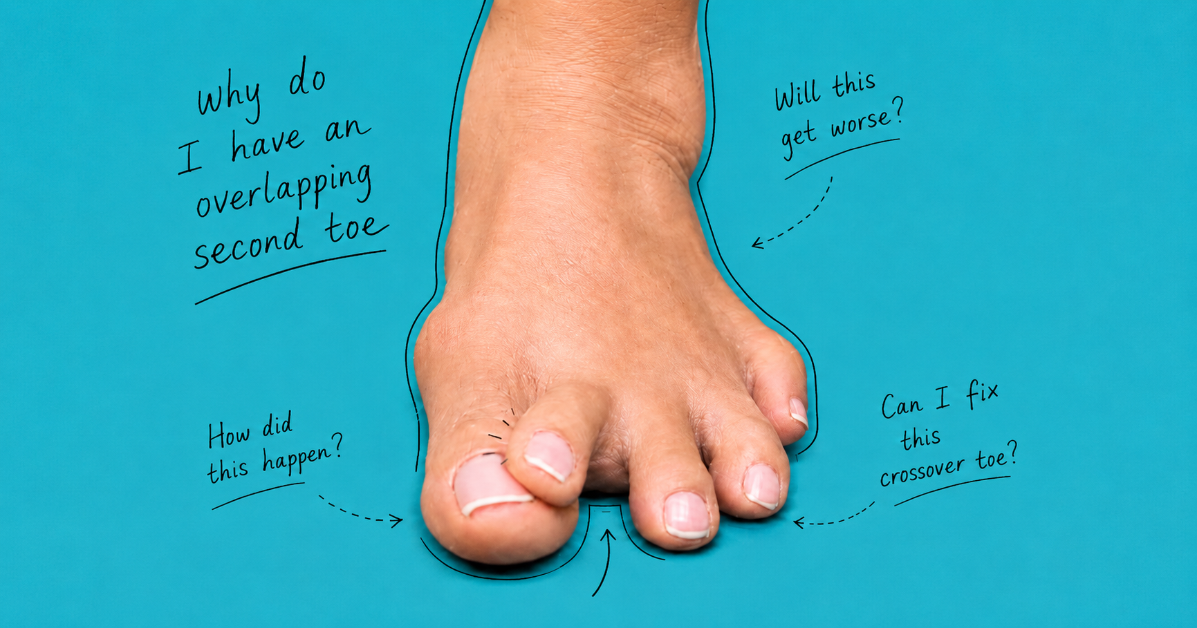
However, plantar plate degeneration can also develop with little or no pain. A new gap between the toes, loss of normal toe contact with the ground, swelling or gradual sideways drift may therefore be important early warning signs. Detecting these changes before the toe becomes obviously elevated, hammered or crossed over may allow the underlying mechanical problem to be addressed earlier
Plantar Plate Degeneration: Why the Joint Beneath Your Toe Starts to Fail
What Is the Plantar Plate?
- the proximal phalanx;
- plantar fascia;
- collateral ligaments;
- deep transverse metatarsal ligament;
- flexor tendon sheath; and
- surrounding capsular structures.
What Does the Plantar Plate Actually Do?
Why “Plantar Plate Degeneration” May Be More Useful Than Simply Saying “Tear”
↓
attenuated
↓
structurally abnormal
↓
partially torn
↓
extensively torn
rather than going from perfectly healthy to completely ruptured in a single event.
Why Is the Second Toe Usually the Problem?
- repetitive second-ray loading;
- forefoot architecture;
- relative metatarsal length;
- hallux valgus;
- altered first-ray function;
- footwear;
- high activity levels; and
- changes in joint stability with age.
The Earliest Warning Sign May Be an Ache Under the Second Toe
- a vague ache;
- a bruised sensation;
- tenderness;
- mild burning;
- discomfort during push-off;
- pain walking barefoot on hard floors; or
- the sensation of standing on a small pebble.
Plantar Plate Degeneration Can Also Be Silent
Patients vary considerably in their awareness of their feet. A toe may gradually move one or two millimetres over many months without producing a dramatic moment when the patient notices something is wrong. The brain becomes accustomed to the appearance of the foot. Eventually someone looks down and thinks: “Has my second toe always been like that?”
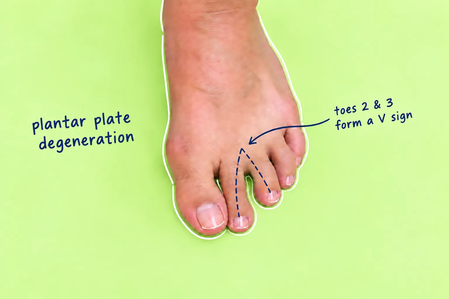
The V-Sign: Look at the Space Between Your Toes
Clinically this is often referred to as a V-sign. But there is an important caveat. Some people naturally have widely spaced toes. Therefore: a V-shaped space is not automatically pathological.
The Toe May Begin Losing Contact With the Ground
- sit slightly higher?
- touch the floor less firmly?
- appear less able to grip?
- feel different during push-off?
Swelling Can Be Another Early Clue
- mild puffiness;
- fullness beneath the joint;
- tenderness;
- discomfort moving the toe; or
- a sensation that one MTP joint feels thicker than the other.
A Simple Home Check
Your Phone May Be Surprisingly Useful
- position;
- camera angle;
- distance;
- lighting; and
- weight-bearing stance.
Do Not Try to Diagnose Instability by Repeatedly Pulling the Toe
From Ache to Crossover Toe: What Progression Can Look Like
“I Feel Like I'm Walking on a Pebble”
This deserves careful assessment because several forefoot conditions can produce remarkably similar symptoms. Possibilities include:
- plantar-plate pathology;
- MTP synovitis;
- capsulitis;
- metatarsalgia;
- Morton neuroma;
- stress injury;
- Freiberg disease;
- intermetatarsal bursitis; and
- other mechanical forefoot problems.
How Is a Plantar Plate Problem Diagnosed?
- precise location of tenderness;
- MTP swelling;
- toe alignment;
- toe purchase;
- drawer testing;
- hallux-valgus assessment;
- metatarsal architecture;
- callus distribution;
- footwear;
- gait and forefoot loading.
Weight-Bearing X-Rays
- metatarsal alignment;
- relative metatarsal lengths;
- hallux valgus;
- MTP subluxation;
- dislocation;
- degenerative joint change; and
- alternative osseous pathology.
Ultrasound
- no radiation;
- relatively low cost;
- dynamic examination;
- ability to examine the structure while moving the toe.
MRI
MRI was more accurate overall and can be particularly useful when determining the extent of injury or assessing associated structures such as collateral ligaments (Albright et al., 2022). But imaging should still be interpreted in context. An MRI finding is not the patient. Symptoms, examination, deformity, stability and imaging should agree before major treatment decisions are made.
Can You Stop Plantar Plate Degeneration From Progressing?
- activity modification;
- footwear changes;
- toe taping or strapping;
- metatarsal offloading;
- orthoses;
- selected rehabilitation;
- calf/Achilles flexibility work where appropriate.
Taping and Strapping
Metatarsal Pads and Orthotics
Footwear
- adequate toe-box width;
- adequate depth;
- cushioning;
- appropriate forefoot stiffness;
- avoidance of excessive heel elevation;
- sufficient shoe length.
What About Exercises?
- maintaining intrinsic-foot function;
- improving toe control;
- maintaining ankle flexibility;
- gradually restoring activity.
Corticosteroid Injections Need Careful Thought
What About PRP?
When Is Surgery Considered?
- persistent pain despite appropriate conservative management;
- progressive instability;
- substantial deformity;
- crossover toe;
- significant subluxation;
- dislocation;
- failure of non-operative treatment.
- direct plantar-plate repair;
- metatarsal osteotomy;
- tendon procedures;
- MTP reconstruction;
- correction of associated hammer toe;
- correction of associated hallux valgus.
Does Plantar Plate Repair Work?
When Should You See a Podiatrist?
- persistent pain beneath the second MTP joint;
- unexplained focal plantar tenderness;
- swelling around the base of a toe;
- a new V-shaped gap;
- progressive toe elevation;
- loss of toe-ground contact;
- sideways drift;
- development of hammering;
- a toe that simply looks different from six months ago.
The Patient We Would Rather See
or even:
“Nothing hurts, but I think this toe is beginning to move.”
The Most Important Message
The Bottom Line
Book An Appointment
Disclaimer: The word "treatment" in this article refers to the care and management of a patient’s health to prevent, cure, or improve a condition. Treatment results vary and do not necessarily indicate a cure. This article is for informational and educational purposes only and does not constitute medical advice.
About the Author
Mark B. Reyneker, BTech (Podiatry), MSc (Palaeontology) is a podiatrist and Founder & Clinical Director of Family Podiatry Centre, with more than 25 years of clinical experience across South Africa, Malaysia and Singapore. His clinical interests include foot and lower-limb pain, gait and biomechanics, sports-related foot conditions, orthotic therapy and footwear.
Alongside his clinical practice, Mark conducts research into human gait and foot biomechanics. His MSc research at the University of the Witwatersrand investigated human propulsion and the structural properties of the metatarsals. He is also the inventor of A Foot Orthotic, an orthotic technology developed through an international patent family.
References
- Albright, R.H., Brooks, B.M., Chingre, M., Klein, E.E., Weil, L.S. Jr and Fleischer, A.E. (2022) ‘Diagnostic accuracy of magnetic resonance imaging (MRI) versus dynamic ultrasound for plantar plate injuries: A systematic review and meta-analysis’, European Journal of Radiology, 152, 110315.
Original article: https://doi.org/10.1016/j.ejrad.2022.110315 - Baker, J.R., Albright, R., Jameson, R., Chingre, M., Klein, E.E., Weil, L.S. Jr and Fleischer, A.E. (2022) ‘Treatment of Lesser Metatarsophalangeal Joint Instability With Plantar Plate Repair: A Systematic Review and Meta-Analysis’, Journal of Foot and Ankle Surgery, 61(5), pp. 1114–1118.
Original article: https://doi.org/10.1053/j.jfas.2022.02.002 - Coughlin, M.J., Schutt, S.A., Hirose, C.B., Kennedy, M.J., Grebing, B.R., Smith, B.W., Cooper, M.T., Golanó, P., Viladot, R. and Alvarez, F. (2012) ‘Metatarsophalangeal Joint Pathology in Crossover Second Toe Deformity: A Cadaveric Study’, Foot & Ankle International, 33(2), pp. 133–140.
Original article: https://doi.org/10.3113/FAI.2012.0133 - Deland, J.T., Lee, K.T., Sobel, M. and DiCarlo, E.F. (1995) ‘Anatomy of the plantar plate and its attachments in the lesser metatarsal phalangeal joint’, Foot & Ankle International, 16(8), pp. 480–486.
Original article: https://doi.org/10.1177/107110079501600804 - Doherty, T.S. et al. (2017) ‘A Systematic Review of Plantar Plate Repair in the Management of Lesser Metatarsophalangeal Joint Instability’, Journal of Foot and Ankle Surgery, 56(6), pp. 1244–1248.
Original article: https://doi.org/10.1053/j.jfas.2017.05.039 - Gregg, J., Marks, P., Silberstein, M., Schneider, T. and Kerr, J.B. (2007) ‘Histologic anatomy of the lesser metatarsophalangeal joint plantar plate’, Surgical and Radiologic Anatomy, 29, pp. 141–147.
Original article: https://doi.org/10.1007/s00276-007-0188-2 - Klein, E.E., Weil, L. Jr, Weil, L.S. Sr, Coughlin, M.J. and Knight, J. (2013) ‘Clinical Examination of Plantar Plate Abnormality: A Diagnostic Perspective’, Foot & Ankle International, 34(6).
Original article: https://doi.org/10.1177/1071100712471825 - Maas, N.M., van der Grinten, M., Bramer, W.M. and Kleinrensink, G.J. (2016) ‘Metatarsophalangeal joint stability: a systematic review on the plantar plate of the lesser toes’, Journal of Foot and Ankle Research, 9, 32.
Original article: https://doi.org/10.1186/s13047-016-0165-2 - Shima, H., Togei, K., Hirai, Y., Tanaka, K., Yasuda, T. and Neo, M. (2024) ‘Ultrasonography-Based Quantitative Evaluation of Second Metatarsophalangeal Joint Instability in Female Patients With Hallux Valgus’, Foot & Ankle International, 45(7), pp. 737–745.
Original article: https://doi.org/10.1177/10711007241239335 - Singer, A.D. et al. (2022) ‘Incidental Finding of Plantar Plate Pathology on Routine Magnetic Resonance Imaging of the Foot and Ankle’.
Original article: https://doi.org/10.1177/19386400221118460 - Talusan, P.G. et al. (2026) ‘Lesser Metatarsophalangeal Joint Instability: Nonsurgical Treatment Alternatives’, Clinics in Podiatric Medicine and Surgery.
Original article: https://doi.org/10.1016/j.cpm.2026.02.011
Frequently Asked Questions
The plantar plate is a thick, rubbery ligament located at the base of your lesser toes (most commonly the second toe). It acts as the primary stabilizer keeping your toe grounded. Plantar plate degeneration occurs when repetitive pressure or mechanical strain causes this ligament to wear down, stretch, or tear away from the bone.
Plantar plate pain typically feels like a persistent, throbbing ache or a sharp burning sensation directly on the ball of the foot, right underneath the base of the second or third toe. Patients often describe it as feeling like they are "walking directly on a marble" or stepping on a folded sock.
When the plantar plate ligament weakens or tears completely, it loses its ability to anchor the toe. This causes the toe to lift off the ground, drift sideways (forming a gap between toes, known as the "V-sign"), or cross over the adjacent toe — a condition known as "crossover toe."
While both conditions cause pain in the ball of the foot, Morton's neuroma is an irritated nerve that typically causes numbness, tingling, or radiating pain between the toes. A plantar plate tear is a ligament injury characterized by localized pinpoint swelling beneath the joint, pain when bending the toe upward, and visible toe drifting or deformity.
The primary cause is repetitive mechanical overload on the forefoot. Common risk factors include overpronation (flat feet), having a second toe that is longer than the big toe (Morton's toe), high arches, bunions, wearing narrow or high-heeled shoes, and high-impact sports like running or tennis.
Yes. High heels force your body weight forward onto the delicate metatarsal heads in the ball of the foot. This extreme angle places severe, continuous stretch on the plantar plate ligament, accelerating wear and tearing over time.
Frequently, yes. A bunion causes the big toe to lean inward, forcing it to lose its ability to bear load during walking. To compensate, the foot shifts excessive pressure onto the adjacent second toe, straining and eventually degenerating its plantar plate.
A podiatrist diagnoses plantar plate injuries through a physical clinical evaluation, including the "Lachman test" (a gentle vertical displacement test of the toe joint), 3D gait analysis to assess pressure points, and imaging such as ultrasound or MRI to confirm the extent of the ligament tear.
Because the plantar plate is soft ligament tissue, it does not show up directly on standard X-rays. However, X-rays are useful to rule out stress fractures, assess joint alignment, and identify toe drifting or joint space narrowing associated with tears.
Yes! Mild to moderate plantar plate tears and degeneration respond very well to non-surgical podiatric care. Conservative treatment focuses on stabilizing the toe joint, reducing strain on the ligament, and correcting underlying foot mechanics so the tissue can heal.
Medical-grade custom orthotics are engineered with specialized metatarsal pads or cutouts. These features redistribute impact weight away from the painful joint head, prevent hyper-extension of the toe during push-off, and stabilize the midfoot arch.
Plantar plate taping (often using "buddy taping" or crossover taping techniques) gently holds the affected toe in a downward, neutral position. This prevents the toe from bending upward during walking, taking tension off the healing ligament.
Yes. Extracorporeal Shockwave Therapy (ESWT) delivers acoustic energy to the chronic tissue under the ball of the foot. This stimulates local blood flow, accelerates collagen repair in degenerate ligament fibers, and provides non-invasive pain relief.
With proper offloading, custom orthotics, and activity modification, conservative recovery usually takes 6 to 12 weeks. Chronic degeneration that has progressed over years may take longer to fully stabilize.
Look for shoes with a wide, roomy toe box (to prevent pinching), low heel-to-toe drop, stiff midsoles, and a gentle rocker sole. Rocker soles allow your foot to roll smoothly through a stride without requiring the toe joints to bend forcefully.
Ice can temporarily numb acute pain after prolonged walking, but it does not heal the ligament. Long-term relief requires structural offloading, toe stabilization, and professional biomechanical correction rather than just cold therapy.
High-impact activities like running, jumping, or HIIT workouts should be temporarily modified or paused while the ligament is healing. Low-impact alternatives such as swimming or cycling in a stiff-soled shoe are recommended until your podiatrist clears you.
If ignored, continuous pressure will cause a partial tear to become a complete rupture. This leads to permanent toe dislocation ("hammer toe" or "crossover toe"), painful bone-on-bone arthritis in the metatarsal joint, and intractable forefoot pain.
Surgery is generally reserved as a last resort for severe, complete ruptures or fixed toe dislocations that failed conservative treatment. The vast majority of early-to-mid-stage cases can be managed non-surgically with podiatric intervention.
Prevent re-injury by wearing supportive, wide-toed footwear, using prescribed custom orthotics to correct faulty gait mechanics, avoiding bare feet on hard surfaces, and addressing secondary issues like bunions or calf tightness early.