Published: 31 August 2026 | Last reviewed: 16 September 2026
Onychophosis is a painful build-up of hard skin around or beneath the edge of a toenail. It can feel remarkably similar to an ingrown toenail, yet the nail may not actually have penetrated the skin. Understanding why this hard skin develops is important because simply cutting the nail or removing the callus may provide temporary relief without addressing the mechanical forces causing it to return.
A painful corner of a toenail is commonly assumed to be an ingrown nail. Sometimes it is. But there is another condition that can produce remarkably similar symptoms: onychophosis.
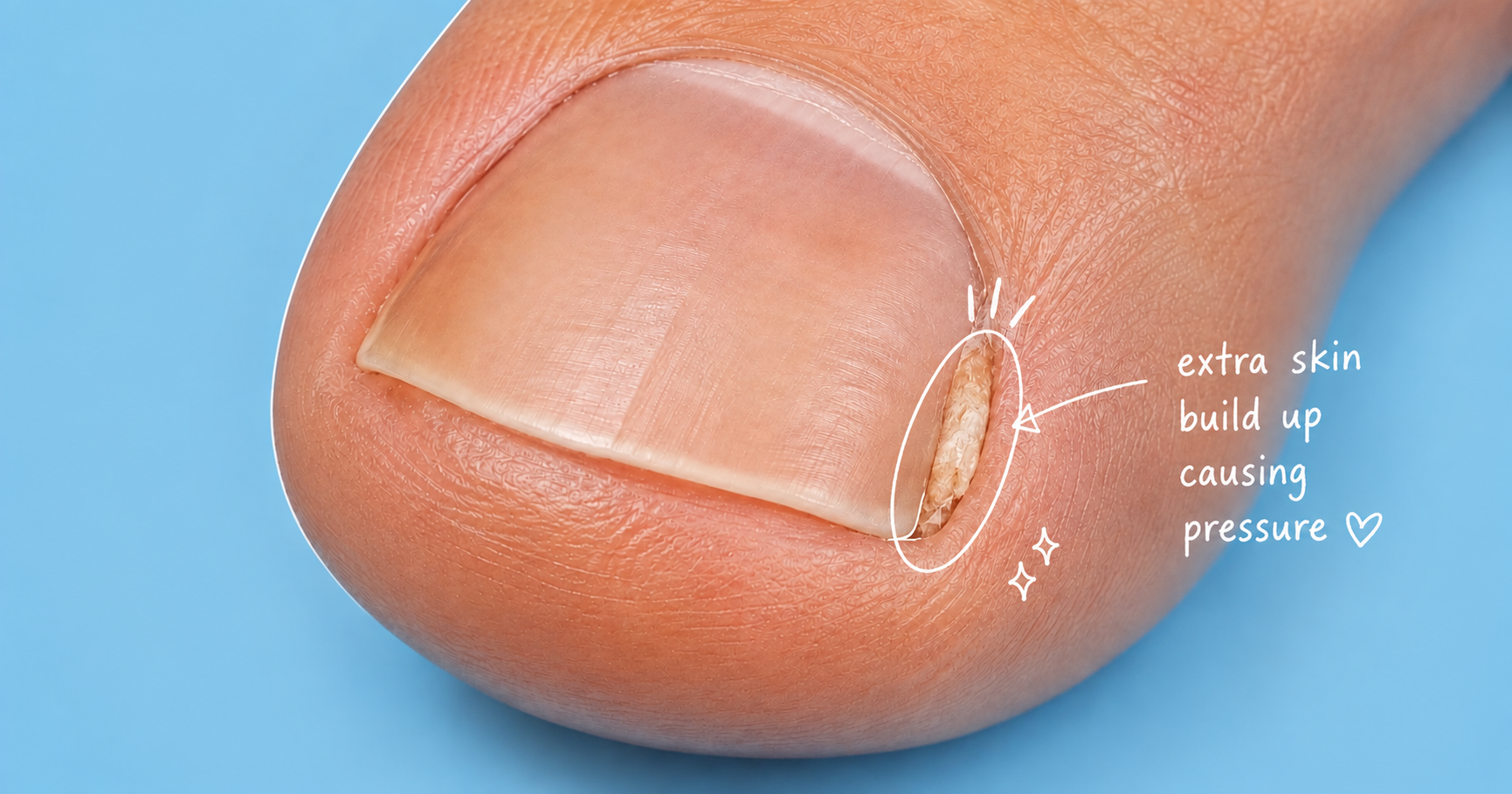
Onychophosis is an accumulation of hyperkeratotic tissue—essentially excessively thickened skin—within the groove beside the nail, around the nail fold or occasionally beneath the nail. It can become extremely painful when compressed between the relatively rigid nail plate and the surrounding tissues. Although podiatrists encounter this condition regularly, onychophosis has received surprisingly little attention as an independent disorder in clinical research.
Much of what we understand about it comes from specialist nail literature and from research into related conditions such as ingrown toenails, mechanical nail disorders and hyperkeratosis. Recent research, however, is increasingly recognising an important principle: many toenail problems are mechanical problems as much as they are skin or nail problems.
A major clinical review published in the Journal of the American Academy of Dermatology in 2026 identified onychophosis among several nail disorders produced by repetitive mechanical trauma. The authors emphasised the effects of footwear, toe deformity, foot structure and gait-related forces on the nail unit (Ricardo et al., 2026).
Understanding this mechanical relationship helps explain not only what onychophosis is, but also why it hurts, why it often returns and why simply cutting deeper into the corner of the nail can sometimes make the problem worse.
What is onychophosis?
The word onychophosis describes hyperkeratosis involving the nail folds, the space between the nail plate and nail fold, or occasionally the area beneath the nail.
Hyperkeratosis means that the outer layer of the skin, the stratum corneum, has become excessively thick. This is essentially the same protective biological response that produces a callus elsewhere on the foot. If repeated pressure is applied to the ball of the foot, for example, the skin may respond by becoming thicker. Around a toenail the same process can occur, but there is much less room available for the thickened tissue.
A 2024 review of nail disorders describes onychophosis particularly around the great and fifth toenails and identifies trauma, poor foot-to-shoe compatibility, hallux valgus, flexed toes and rotation of the fifth toe as potential contributing factors (Albucker, Conway and Lipner, 2024).
This gives us a simple way of understanding the disorder:
Onychophosis is, in many cases, a pressure-induced callus occurring in a very confined anatomical space. And that confined space is one of the reasons it can hurt so much.
Why can such a small piece of hard skin hurt so much?
A callus across a broad area underneath the foot distributes its pressure across a relatively large surface.
The nail groove is very different. Imagine a small piece of hardened keratin being compressed between:
the edge of the nail plate on one side and the soft tissue of the toe on the other.
Even a relatively small increase in tissue thickness can considerably increase local pressure. People with painful nail disorders may describe pain as sharp, shooting, throbbing or burning, and symptoms can become worse with walking, footwear and direct pressure (Olvera-Rodríguez et al., 2021).
This is why a tiny area beside a toenail can sometimes produce disproportionately severe pain.
Book An Appointment Onychophosis and ingrown toenails are not the same thing
This distinction is particularly important.
An ingrown toenail, medically called onychocryptosis, occurs when the nail edge impinges upon or penetrates the surrounding nail fold. This may result in pain, inflammation, swelling, granulation tissue and sometimes secondary infection (Geizhals and Lipner, 2019; Thakur, Vinay and Haneke, 2020).
In onychophosis, the painful tissue is primarily hyperkeratotic skin. The two conditions can nevertheless coexist—and one may contribute to the development of the other.
Haneke (2012) described an interesting sequence in which pressure from a nail edge initially produces a painful localised area of hyperkeratosis: onychophosis. The person then tries to relieve the discomfort by repeatedly cutting further down the corner of the nail.
Unfortunately, this can leave a sharp nail spicule. As that nail grows forward, the spicule may penetrate the nail fold and produce genuine onychocryptosis.
The sequence may therefore look something like:
Nail pressure
↓
Painful hyperkeratosis / onychophosis
↓
Repeated cutting down the nail corner
↓
Nail spicule
↓
Penetration of the nail fold
↓
Inflammation and an ingrown toenail
This does not mean that every case of onychophosis will become an ingrown nail.
It does explain, however, why aggressively cutting down the sides of a painful nail can occasionally transform one problem into another.
Why does onychophosis develop?
The most useful way to think about onychophosis is not simply as excessive skin growth, but as the skin's response to repeated mechanical stress. The skin does not randomly decide to produce callus. Keratinisation is protective.
When an area is repeatedly exposed to pressure or friction, the epidermis can increase production of keratinised tissue to protect itself. Around the toenail, however, this protective response can become counterproductive.
As the tissue becomes thicker, the available space between the nail and surrounding skin decreases. The additional material can then increase local pressure, which may stimulate further irritation.
A potential cycle develops:
Pressure or friction
↓
Repeated microtrauma
↓
Increased keratinisation
↓
Thicker tissue in the nail groove
↓
Greater compression
↓
Pain and further mechanical irritation
This also helps explain one of the most frustrating features of onychophosis:
Why does it keep coming back?
Removing the hard skin can reduce the pressure and often provides considerable relief. But removing the keratin does not necessarily remove the reason the keratin developed. If the same mechanical forces continue acting on the nail groove, the skin may simply produce more protective keratin.
In other words:
Debridement removes the consequence.
Long-term management should also consider the cause.
The role of shoes
Tight shoes are commonly blamed for nail problems, but describing footwear simply as "too tight" is probably an oversimplification.bA more useful concept is foot-to-shoe incompatibility.
A shoe may technically be the correct size but still place excessive pressure against one particular toe because of its:
- toe-box shape
- toe-box width
- toe-box depth
- upper material
- taper
- internal seams
- interaction with the person's particular toe alignment.
The 2026 review by Ricardo et al. identifies inappropriate footwear as an important contributor to mechanically induced nail pathology and emphasises examining the relationship between the foot, toes and footwear rather than examining the nail in isolation (Ricardo et al., 2026).
For example, a person with a rotated fifth toe may repeatedly press the lateral aspect of that nail against the shoe even when the shoe does not feel particularly small.
Similarly, hallux valgus can alter the relationship between the great toe and second toe and potentially create compression around one side of the nail.
This may explain why the great toe and fifth toe are particularly common sites of onychophosis (Albucker, Conway and Lipner, 2024).
Toe shape and foot mechanics matter too
Modern nail literature is increasingly recognising that the nail should not be examined independently of the rest of the foot.
Mechanical and structural conditions associated with nail abnormalities include:
- hallux valgus
- hallux rigidus
- hallux erectus
- flexed or hammer toes
- rotated fifth toes
- transverse nail overcurvature
- pincer nails
- altered forefoot structure
- repetitive trauma from footwear or activity.
Ricardo et al. (2026) argue that dystrophic toenails should be assessed together with toe and foot alignment during both standing and walking.
This is an important shift in thinking.
A painful nail may be the site where the problem becomes visible, while the force producing the problem originates elsewhere.
For example:
toe rotation → shoe pressure → nail-fold compression → hyperkeratosis
or
curved nail → nail-fold pressure → hyperkeratosis
or
adjacent toe pressure → lateral nail compression → hyperkeratosis
Different mechanisms can therefore produce a very similar-looking lesion.
Nail curvature may contribute
Some nails become increasingly curved across their width. At the extreme end of the spectrum is a pincer nail, where the sides of the nail curve downward and may compress the nail bed or nail folds. Pincer nails can cause substantial pain and are influenced by both anatomical and mechanical factors (Huang et al., 2020).
Haneke (2012) also describes older patients in whom increasingly curved great toenails exert pressure upon the nail groove, producing onychophosis even before the nail actually breaks the skin.
In these cases, repeatedly removing the hard skin may provide relief, but the curvature of the nail continues generating the pressure. This explains why treatment sometimes needs to address both the skin and the nail.
Is onychophosis a fungal infection?
Not necessarily. This is another important distinction.
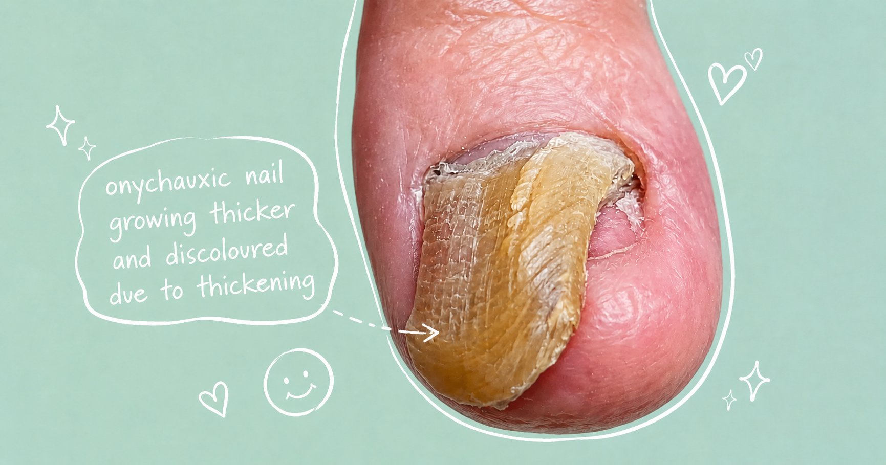
Fungal nail infection—or onychomycosis—can cause:
- nail thickening
- nail discolouration
- crumbling
- separation of the nail
- subungual debris
- subungual hyperkeratosis.
Mechanical nail damage can produce several of the same changes.
Recent research increasingly warns against diagnosing fungal nail disease purely by appearance.
In a study of patients with transverse nail overcurvature and subungual hyperkeratosis, only 28 of 65 samples—43.1%—were positive for onychomycosis. More than half therefore had the characteristic hyperkeratotic appearance without confirmed fungal infection (Di Chiacchio et al., 2021).
Similarly, research into foot and toe deformity suggests that repetitive mechanical trauma may damage the nail unit and can also predispose the nail to secondary fungal infection (Haneke, 2024). So mechanical nail disease and fungal nail disease are not necessarily competing diagnoses.
A nail can be:
mechanically damaged without fungus, infected with fungus, or mechanically damaged and subsequently infected with fungus.
When fungal infection is suspected, laboratory confirmation may therefore be appropriate before committing someone to prolonged antifungal treatment.
What about a corn underneath the nail?
Another condition that can resemble onychophosis is an onychoclavus, sometimes called a subungual heloma. This is essentially a more discrete corn-like hyperkeratotic lesion beneath the nail. Onychophosis tends to describe hyperkeratotic tissue around the nail fold, within the nail groove or sometimes subungually, whereas onychoclavus refers more specifically to a focal subungual corn.
For a patient, a useful simplified distinction is:
Onychophosis = nail-groove callus
Onychoclavus = corn underneath the nail
Both may have a mechanical origin and both can be extremely painful because the hardened tissue is compressed in a confined space (Albucker, Conway and Lipner, 2024).
How is onychophosis diagnosed?
Diagnosis is usually clinical. The important part of the examination, however, is not simply identifying the hard skin. The clinician should try to determine why it formed.
An examination may therefore include:
1. The nail
- Is it unusually curved?
- Is it thickened?
- Has it been cut deeply down the sides?
- Is there a sharp nail spicule?
- Is the nail actually penetrating the skin?
- Is there subungual debris?
2. The nail groove
- Is the painful structure hard keratin?
- Is there inflammation?
- Is there granulation tissue?
- Is there discharge or infection?
3. The toe
- Is the great toe deviated?
- Is the fifth toe rotated?
- Is another toe pressing against the affected nail?
- Is the toe flexed or hammered?
4. The shoe
- Where does the shoe place pressure?
- Does the toe box taper?
- Is there sufficient width and depth?
- Does the foot slide forward inside the shoe?
5. The foot
Where relevant, standing and walking assessment may help determine whether toe position or forefoot mechanics contribute to repetitive nail trauma. This broader examination is consistent with the emerging concept that mechanically induced nail disorders should be assessed together with foot structure and gait rather than treating the nail as an isolated structure (Ricardo et al., 2026).
How is onychophosis treated?
The evidence specifically studying treatments for onychophosis is limited. This is worth stating clearly. There are good clinical descriptions and expert reviews, but there are not large randomised controlled trials comparing different onychophosis treatments. Treatment recommendations therefore combine specialist nail literature with evidence from related hyperkeratotic and mechanical conditions.
1. Remove the painful hyperkeratosis
Careful debridement of the accumulated keratin reduces the volume of tissue trapped within the nail groove. This can substantially reduce local pressure and also allows the clinician to see the underlying nail edge more clearly. Debridement is specifically recommended in contemporary reviews of onychophosis (Albucker, Conway and Lipner, 2024).
For many patients, this produces rapid symptomatic improvement. However, recurrence should not automatically be interpreted as treatment failure. If the mechanical stimulus continues, the skin may respond by producing keratin again.
2. Reduce pressure
This is arguably the most important longer-term principle.
Depending upon the cause, pressure reduction might involve:
- changing shoe shape
- providing greater toe-box width or depth
- reducing pressure from an adjacent toe
- accommodating a rotated toe
- addressing repetitive activity-related pressure
- modifying the way the nail is cut
- managing excessive nail thickness or curvature.
The objective is not simply to make the shoe "bigger". It is to identify where the pressure is occurring and why.
3. Keratolytic treatment may sometimes help
Keratolytic agents soften or break down excessive keratin.
A contemporary review lists treatments including approximately:
- urea 20%
- ammonium lactate 12%
- salicylic acid 6–20%
for onychophosis (Albucker, Conway and Lipner, 2024). However, an important evidence distinction is necessary.
These recommendations are mainly based on clinical experience and broader dermatological evidence rather than trials conducted specifically on onychophosis. Randomised studies of hyperkeratotic and dry foot skin demonstrate that urea-, salicylic-acid- and ammonium-lactate-containing preparations can reduce abnormal scaling and improve skin condition (Jennings et al., 1998; Ademola et al., 2002).
That provides biological support for their use, but it does not establish an ideal drug, concentration or treatment schedule specifically for onychophosis. Strong keratolytics should also be used with particular caution around compromised skin and in people with conditions affecting circulation or sensation.
4. Sometimes the nail itself needs treatment
If the principal problem is excessive nail curvature or repeated pressure from the nail edge, treating only the hard skin may not be sufficient.
Conservative approaches to nail curvature and early ingrowing nails may include appropriate nail cutting, packing or gutter techniques and, in selected cases, nail-bracing techniques. If there is genuine recurrent onychocryptosis, partial nail surgery may eventually become appropriate.
But it is important not to confuse the evidence for ingrown toenail surgery with evidence for isolated onychophosis. A 2023 systematic review of randomised trials found that adding phenolisation of the nail matrix after nail avulsion appeared to reduce recurrence, although the authors emphasised that the overall quality of the available evidence was poor (Exley et al., 2023).
More recently, a 2026 systematic review and network meta-analysis involving 44 studies and 5,068 participants found that simple nail avulsion had the greatest recurrence risk. Chemical matricectomy, minor surgery, conventional surgery and conservative treatments showed less clear differences from one another, while conservative approaches generally produced less pain and faster recovery (Morioka et al., 2026).
The practical message is therefore not:
"Recurrent nail pain requires surgery."
It is:
Correctly identify what is producing the pain and match the treatment to that pathology.
Why repeatedly digging down the nail can make things worse
People suffering from onychophosis often discover that cutting down the side of the nail provides temporary relief. This is understandable. Removing part of the nail may temporarily create more space.
The problem is that repeated deep cutting can leave an irregular or sharp edge. As the nail subsequently grows forward, that edge may behave like a small spike and traumatise the nail fold.
Haneke described precisely this cycle in the development of ingrown toenails (Haneke, 2012). Persistent painful nail corners are therefore better assessed than repeatedly excavated at home.
When painful hard skin around a nail needs further investigation
Most onychophosis is benign. However, persistent nail lesions should not automatically be assumed to be callus, fungus or an ingrown nail.
A subungual exostosis, for example, is a benign bony growth arising from the distal phalanx that can produce pain and nail deformity. Radiographs are particularly useful when this is suspected (DaCambra, Gupta and Ferri-de-Barros, 2014; Pérez-Palma, Manzanares-Céspedes and Giralt de Veciana, 2018).
An onychopapilloma, a benign nail-bed tumour, can produce a small keratotic mass beneath the distal nail, often associated with a longitudinal nail change or distal fissure (Tosti et al., 2016).
Rarely, malignant nail disease can also masquerade as persistent benign nail pathology. Nail-unit squamous cell carcinoma can produce hyperkeratosis, nail destruction, ulceration and other relatively nonspecific features, which is one reason diagnosis may be delayed (Tordjman et al., 2025; Venturi et al., 2025).
Further assessment is particularly appropriate when a lesion:
- persists despite appropriate treatment
- repeatedly bleeds
- ulcerates
- produces unexplained progressive nail destruction
- changes significantly in appearance
- develops unusual pigmentation
- produces persistent unexplained pain
- behaves differently from an ordinary recurrent corn or nail lesion.
This does not mean that these symptoms usually indicate something serious.
It means that persistent nail disease deserves a diagnosis rather than repeated assumptions.
The bigger lesson: the nail is part of the foot
Perhaps the most interesting development in modern nail research is the increasing recognition that toenails are mechanically influenced structures. They sit at the end of a moving limb, inside footwear, surrounded by other toes and exposed to thousands of loading cycles during walking and exercise.
Toe position, nail curvature, footwear and repetitive loading can therefore interact. This means that what appears to be a small skin problem beside the toenail may actually represent the final stage of a much broader mechanical process.
With onychophosis, the visible hard skin may be the body's response. The underlying problem may be the force that caused the skin to respond.
A useful way of thinking about recurrent onychophosis is therefore:
NAIL SHAPE × TOE POSITION × FOOTWEAR × REPETITIVE PRESSURE
↓
NAIL-FOLD COMPRESSION
↓
SKIN ADAPTATION
↓
HYPERKERATOSIS
↓
PAIN
This also explains why removing the hard skin can make someone feel considerably better while not necessarily preventing it from returning.
For longer-term management, the question becomes not only:
"How do we remove the painful skin?"
but also:
"Why is this particular area being repeatedly compressed?"
That distinction can make the difference between repeatedly treating the symptom and understanding the mechanism producing it.
Book An Appointment
Disclaimer: The word "treatment" in this article refers to the care and management of a patient’s health to prevent, cure, or improve a condition. Treatment results vary and do not necessarily indicate a cure. This article is for informational and educational purposes only and does not constitute medical advice.
About the Author
Mark B. Reyneker, BTech (Podiatry), MSc (Palaeontology) is a podiatrist and Founder & Clinical Director of Family Podiatry Centre, with more than 25 years of clinical experience across South Africa, Malaysia and Singapore. His clinical interests include foot and lower-limb pain, gait and biomechanics, sports-related foot conditions, orthotic therapy and footwear.
Alongside his clinical practice, Mark conducts research into human gait and foot biomechanics. His MSc research at the University of the Witwatersrand investigated human propulsion and the structural properties of the metatarsals. He is also the inventor of
A Foot Orthotic, an orthotic technology developed through an international patent family.
References
1. Ademola, J., Frazier, C., Kim, S.J., Theaux, C. and Saudez, X. (2002) ‘Clinical evaluation of 40% urea and 12% ammonium lactate in the treatment of xerosis’,
American Journal of Clinical Dermatology, 3(3), pp. 217–222. doi: 10.2165/00128071-200203030-00007.
Original article / DOI3. DaCambra, M.P., Gupta, S.K. and Ferri-de-Barros, F. (2014) ‘Subungual exostosis of the toes: a systematic review’,
Clinical Orthopaedics and Related Research, 472(4), pp. 1251–1259. doi: 10.1007/s11999-013-3345-4.
Original article / DOIFree full text on PubMed Central4. Exley, V., Jones, K., O’Carroll, G., Watson, J. and Backhouse, M. (2023) ‘A systematic review and meta-analysis of randomised controlled trials on surgical treatments for ingrown toenails part I: recurrence and relief of symptoms’,
Journal of Foot and Ankle Research, 16, 35. doi: 10.1186/s13047-023-00631-1.
Original article / DOIFree full text on PubMed Central5. Geizhals, S. and Lipner, S.R. (2019) ‘Review of onychocryptosis: epidemiology, pathogenesis, risk factors, diagnosis and treatment’,
Dermatology Online Journal, 25(9). doi: 10.5070/D3259045583.
Original full-text articleDOI8. Huang, C., Huang, R., Yu, M., Guo, W., Zhao, Y., Li, R. and Zhu, Z. (2020) ‘Pincer nail deformity: clinical characteristics, causes, and managements’,
BioMed Research International, 2020, 2939850. doi: 10.1155/2020/2939850.
Original article / DOIFree full text on PubMed Central9. Jennings, M.B., Alfieri, D., Ward, K. and Lesczczynski, C. (1998) ‘Comparison of salicylic acid and urea versus ammonium lactate for the treatment of foot xerosis: a randomized, double-blind, clinical study’,
Journal of the American Podiatric Medical Association, 88(7), pp. 332–336. doi: 10.7547/87507315-88-7-332.
Original article / DOIPubMed record10. Morioka, D., Matsuoka, Y., Tamagawa, K., Takagi, S., Tomizuka, Y., Okada, N. and Tamura, S. (2026) ‘Comparative effectiveness of ingrown toenail treatments: systematic review and network meta-analysis’,
Journal of Foot and Ankle Surgery, published online 24 August 2026. doi: 10.1053/j.jfas.2026.08.021.
Original article / DOIPubMed record11. Noriega, L.F., Di Chiacchio, N.G., de Mello, C.D.B.F.
et al. (2020) ‘Prevalence of onychomycosis among patients with transverse overcurvature of the nail: results of a cross-sectional study’,
Skin Appendage Disorders, 6(6), pp. 351–354. doi: 10.1159/000509985.
Original article / DOIFree full text on PubMed Central12. Olvera-Rodríguez, V., Gatica-Torres, M., Carrillo-Córdova, D.M., Barrera-Godínez, A. and Domínguez-Cherit, J. (2021) ‘Painful nails: a practical approach to the diagnosis and management of painful nail conditions’,
International Journal of Dermatology, 60(11), pp. 1318–1333. doi: 10.1111/ijd.15496.
Original article / DOIPubMed record13. Pérez-Palma, L., Manzanares-Céspedes, M.C. and Giralt de Veciana, E. (2018) ‘Subungual exostosis: systematic review and meta-analysis’,
Journal of the American Podiatric Medical Association, 108(4), pp. 320–333. doi: 10.7547/17-102.
Original article / DOIPubMed record14. Ricardo, J.W., Dominguez-Cherit, J., Haneke, E., Rana, J., Richert, B., Tosti, A., Vlahovic, T.C. and Lipner, S.R. (2026) ‘Evaluation and management of mechanical and structural nail disorders: a clinical review’,
Journal of the American Academy of Dermatology, 94(5), pp. 1493–1503. doi: 10.1016/j.jaad.2025.11.025.
Original article / DOIPubMed record15. Thakur, V., Vinay, K. and Haneke, E. (2020) ‘Onychocryptosis – decrypting the controversies’,
International Journal of Dermatology, 59(6), pp. 656–669. doi: 10.1111/ijd.14769.
Original article / DOIPubMed record16. Tordjman, L., Warp, P.V., Robinson-Pinkney, C. and Morrison, B. (2026) ‘Evaluating dermoscopic features of nail unit squamous cell carcinoma: a systematic review and sub-analysis’,
Skin Appendage Disorders, 12(3), pp. 171–178. Published online 12 December 2025. doi: 10.1159/000550020.
Original article / DOIPubMed record17. Tosti, A., Schneider, S.L., Ramirez-Quizon, M.N., Zaiac, M. and Miteva, M. (2016) ‘Clinical, dermoscopic, and pathologic features of onychopapilloma: a review of 47 cases’,
Journal of the American Academy of Dermatology, 74(3), pp. 521–526. doi: 10.1016/j.jaad.2015.08.053.
Original article / DOIPubMed record18. Venturi, F., Magnaterra, E., Scotti, B., Alessandrini, A., Veneziano, L., Vaccari, S., Baraldi, C. and Dika, E. (2025) ‘Squamous cell carcinoma of the nail unit: a comprehensive review of clinical features, diagnostic workflow, management strategies and therapeutic options’,
Diagnostics, 15(18), 2378. doi: 10.3390/diagnostics15182378.
Original article / DOIFree full text on PubMed Central