Why Does My Heel Hurt When I First Get Out of Bed? The Science of Early-Morning Heel Pain



19 September 2026
By Mark Reyneker, BTech (Podiatry), MSc (Palaeontology), Registered Podiatrist, Foot & Gait Specialist | Founder & Clinical Director, Family Podiatry Centre
Explainer video
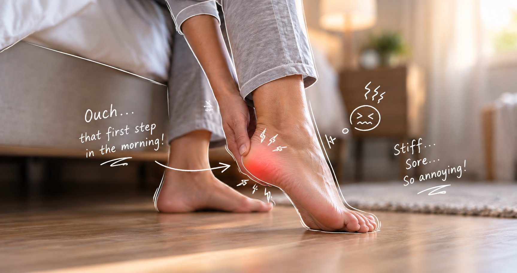
Heel pain with the first few steps in the morning is commonly associated with plantar fasciopathy, often called plantar fasciitis. The pain appears to occur because the plantar heel has spent several hours relatively unloaded and is then suddenly required to accept bodyweight.
Why Does My Heel Hurt When I First Get Out of Bed? The Science of Early-Morning Heel Pain
Then something equally interesting happens. The same thing may occur after sitting through dinner, driving for an hour, watching television or spending a long time at a desk. Stand up again and the heel suddenly reminds you that it is there.
This pattern is often called first-step pain or post-static pain.
It is one of the most characteristic features of plantar fasciopathy—the condition still commonly called plantar fasciitis—and it tells us something fascinating about how connective tissue, mechanical loading and pain interact. But what actually happens during those hours or minutes of rest? And why can a heel that was relatively comfortable while lying in bed suddenly become painful the instant bodyweight passes through it?
The explanation turns out to be more complicated than the familiar idea that the plantar fascia simply “tightens overnight”.
First-step pain is one of the strongest clues in plantar heel pain
The pain is typically most noticeable:
- with the first steps after getting out of bed;
- when standing after prolonged sitting;
- at the beginning of walking after inactivity; and
- sometimes again after prolonged standing, walking or exercise.
The 2023 clinical practice guideline for plantar heel pain identifies plantar-medial heel pain that is most noticeable during the initial steps after inactivity as one of the defining features of plantar fasciopathy (Koc et al., 2023). In a clinical study examining 250 people with plantar heel pain, approximately 88% of those diagnosed with plantar fasciitis reported first-step morning pain (Yi et al., 2011). So the phenomenon is real and remarkably consistent. What is less certain is exactly why it happens.
The popular explanation: the fascia shortens overnight
Healing begins in this shortened position. When the person stands the following morning, the fascia is suddenly stretched back to its functional length and microscopic tissue disruption occurs again. It is an attractive explanation.
It has also been repeated in textbooks, patient information sheets and discussions around night splints for decades. There is probably some truth in the general concept of prolonged unloading followed by abrupt reloading.
However, there is surprisingly little direct experimental evidence showing that a pathological human plantar fascia literally contracts overnight and then repeatedly tears open every morning. The modern explanation is likely more nuanced.
Think about the transition from zero load to bodyweight
Within seconds, approximately your entire bodyweight—and during walking, transiently more than bodyweight—is transferred through the foot. The arch begins to deform. The calcaneus and forefoot move relative to one another. The plantar fascia becomes tensioned. The Achilles tendon becomes loaded. The intrinsic foot muscles become active. The heel fat pad is compressed. The plantar fascial attachment to the calcaneus experiences tension, shear and compression.
A healthy foot handles this transition without complaint. A sensitised plantar heel may not.
The plantar fascia is viscoelastic
That means its mechanical behaviour depends not only upon how much force is applied but also upon:
- how quickly the force is applied;
- how long the force is maintained;
- its previous loading history; and
- the orientation of the collagen fibres.
Pettenuzzo et al. demonstrated that human plantar fascia is strongly anisotropic—its mechanical behaviour depends upon the direction in which it is loaded—and exhibits distinctly nonlinear, time-dependent viscoelastic behaviour. Its collagen fibres are predominantly orientated longitudinally from heel towards forefoot, exactly as we would expect from the forces repeatedly placed through the foot during locomotion (Pettenuzzo et al., 2026).
This matters enormously when thinking about first-step pain. The plantar fascia that receives the first morning step has spent hours in an almost completely different loading environment. It is then subjected to a relatively abrupt mechanical transition.
How far does the plantar fascia actually stretch when we walk?
For an irritated or sensitised plantar fascial attachment, the first sudden exposure to that strain after hours of unloading can produce a very different sensory experience. That is probably closer to what happens when the first foot hits the floor in the morning.
The painful area is usually an enthesis
At the plantar fascial origin, tissue changes progressively from: dense collagen → uncalcified fibrocartilage → calcified fibrocartilage → bone. This transition allows forces to move between a relatively flexible collagen structure and a much stiffer calcaneus. That is mechanically difficult territory.
The plantar fascial enthesis experiences more than simple longitudinal pulling. It is exposed to combinations of tension, compression, bending and shear. Fibrocartilage at the plantar fascial attachment appears specifically adapted to these complex forces. This may also help explain why plantar fasciopathy so commonly develops close to the medial calcaneal attachment rather than randomly halfway along the fascia.
Now imagine the first step
As walking begins, the toes progressively dorsiflex and the windlass mechanism increases plantar fascial tension further. At the same time, the Achilles tendon is transmitting force through the posterior calcaneus. Intrinsic foot muscles become increasingly active. The heel fat pad compresses against the floor.
The plantar fascial enthesis suddenly moves from a low-load environment to a complex multi-directional loading environment. If that enthesis and the surrounding tissues are mechanically sensitive, the nervous system receives a powerful burst of nociceptive input. And you feel that first step.
Is the fascia actually tearing again every morning?
Histological studies of chronic plantar fasciopathy demonstrate collagen degeneration, myxoid change and tissue remodelling (Lemont, Ammirati and Usen, 2003). But there is no convincing evidence that every painful morning step creates a fresh structural tear. If that were happening on a meaningful scale every morning, we would expect a rather different injury pattern. The pain can instead be understood as a sensitive tissue responding strongly to rapid mechanical loading. That distinction is important. Pain does not necessarily mean that tissue is being freshly damaged each time it hurts.
The nerves appear to matter considerably
In one study comparing people with unilateral plantar heel pain with healthy controls, patients demonstrated widespread reductions in pressure-pain thresholds—not just beneath the painful heel but at other musculoskeletal and nerve locations. Most interestingly, increased sensitivity of lower-limb nerve trunks was associated with greater first-step morning pain (Fernández-Lao et al., 2019).
Another study mapping pressure sensitivity across the feet found bilateral hypersensitivity even in people who reported symptoms in only one heel. The degree of sensitivity was also associated with morning first-step pain (Saban et al., 2019). This gives us a much more complete model. The first step is mechanically demanding. But the amount of pain generated by that step also depends upon how sensitive the local and central nervous systems have become to that mechanical input.
Chronic heel pain may gradually become a different pain problem
Nevertheless, it adds to a growing body of evidence suggesting that persistent plantar heel pain may eventually involve more than local connective-tissue pathology alone (Karakılıç, Selçuk and Öztürk, 2025). This might help explain something podiatrists see regularly: two patients can have plantar fasciae that look similarly abnormal on ultrasound, yet one experiences relatively mild symptoms while the other experiences severe first-step pain.
What happens during the hours of rest?
1. Mechanical unloading
2. The foot often rests in a relatively plantarflexed position
This provides the mechanical rationale for night splints, which attempt to keep the ankle in dorsiflexion rather than allowing prolonged plantarflexion. However, whether this prevents literal “shortening” of the fascia is less certain than is sometimes suggested.
3. The intrinsic foot muscles are inactive
4. A sensitised enthesis remains sensitised
Why does sitting for 30 minutes reproduce the same problem?
The same phenomenon can occur:
- after sitting through lunch
- after a long car journey
- after working at a computer
- after watching a film
That is why I find the term post-static pain particularly useful. The tissues have been relatively static and unloaded. Then they are suddenly required to accept bodyweight and participate in locomotion again.
Why does the pain often disappear after walking?
This is sometimes called the warm-up phenomenon. It occurs in several load-related connective-tissue disorders, including Achilles tendinopathy. “Warming up” is a useful description of what the patient feels, but actual temperature change is unlikely to explain the entire phenomenon. Several things happen simultaneously.
The tissue's loading state changes
The muscles begin sharing the load
The nervous system also adapts to repeated input
Then why can the heel hurt again later in the day?
The patient can therefore feel much better at 10 am than at 7 am, and then considerably worse again by 6 pm. This pattern makes sense when plantar fasciopathy is viewed as a load-capacity problem rather than simply an inflammatory condition.
Morning “stiffness” and morning “pain” are not necessarily identical
Others describe the foot as:
- stiff;
- tight;
- wooden;
- reluctant to move;
- difficult to flatten;
- or uncomfortable rather than painful.
The distinction can be useful. A sensation of stiffness does not necessarily mean the plantar fascia has physically become shorter. Perceived stiffness is influenced by connective-tissue mechanics, joint motion, muscle tone and sensory processing. This is well recognised in tendon disorders. Achilles tendinopathy, for example, frequently produces morning stiffness that improves after several minutes of walking. The plantar heel may behave in a similar way.
Is morning pain related to how thick the plantar fascia is?
A 2022 study following 90 patients found that baseline ultrasound findings did not predict clinical outcome particularly well. Reductions in plantar fascia thickness over six months were associated with improvements in symptoms, including morning pain, but the correlations were relatively modest (Johannsen et al., 2022). This fits with the broader picture. Structure matters. But structure alone is not pain.
Does stretching before getting out of bed make sense?
Recent biomechanical work gives us additional insight. A 2026 cadaveric investigation measured strain directly within the plantar fascia during different stretching techniques. Dorsiflexion of the toes combined with targeted pressure against the plantar aponeurosis produced measurable fascial strain more consistently than toe dorsiflexion alone (Nazarian et al., 2026).
So gently loading and moving the plantar fascia before asking it to suddenly accept full bodyweight has a reasonable mechanical basis. It is not necessarily “breaking up scar tissue”. It is preparing a load-sensitive structure for the transition from rest to standing.
What about night splints?
The 2023 American physical therapy guideline continues to recommend a one-to-three-month programme of night splints for people whose plantar heel pain is consistently worst with their first morning steps. Singapore's clinical consensus guideline similarly considers night splints a possible adjunct when symptoms persist despite initial management. The evidence is not perfect.
Randomised studies have not consistently demonstrated large additional benefits when night splints are added to otherwise comprehensive rehabilitation programmes. Their usefulness probably varies considerably between individuals.
Morning heel pain does not automatically mean plantar fasciitis
Plantar fasciopathy
Heel fat-pad pain
Achilles tendinopathy
Nerve-related heel pain
Calcaneal stress injury
Inflammatory enthesitis
Persistent bilateral heel pain, prolonged morning stiffness, other joint pain, inflammatory back symptoms, psoriasis, inflammatory bowel disease or a history of uveitis changes the clinical picture considerably. In that situation, the heel may be telling us something about a systemic inflammatory disorder rather than simply local mechanical overload.
Why I find first-step pain so clinically useful
There is a difference between:
- a heel that hurts immediately after rest but improves after walking;
- a heel that becomes progressively worse with every kilometre;
- a heel that feels bruised whenever it strikes a hard surface;
- a heel that burns or tingles;
- and a heel that remains painful at night while the patient is lying in bed.
They may all be described initially as “heel pain”. Biologically, however, they can represent quite different problems. First-step pain gives us an unusually useful clue because it tells us how the tissue responds to a very specific event: the transition from unloading to loading.
What I think is actually happening during that first morning step
If plantar fasciopathy is present, the proximal fascia and its calcaneal enthesis may already contain altered collagen, extracellular-matrix changes and abnormal mechanical properties. The local nociceptive system may also be sensitised.
Then the person stands. Within seconds the arch deforms under bodyweight. The plantar fascia becomes tensioned. The enthesis receives tensile, compressive and shear forces. The Achilles and intrinsic muscles begin working. The heel pad compresses. The first step therefore represents an abrupt transition from almost no functional load to a complex, rapidly applied load.
That is why the phenomenon occurs after lunch, a car journey or an evening on the sofa as well as after eight hours in bed.
The first-step phenomenon tells us something larger about plantar fasciopathy
The condition appears to involve interactions between:
- mechanical load;
- collagen and extracellular-matrix adaptation;
- the plantar fascial enthesis;
- foot and calf mechanics;
- muscle function;
- local nociception;
and, in some chronic cases, altered pain processing. Early-morning heel pain captures all of these processes remarkably well. It is not simply a stiff band underneath the foot. It is what happens when a mechanically specialised, biologically active and sometimes sensitised load-bearing system is suddenly asked to go from rest to work. And that may explain why those first five steps can feel so different from the next fifty.
Book An Appointment
Disclaimer: The word "treatment" in this article refers to the care and management of a patient’s health to prevent, cure, or improve a condition. Treatment results vary and do not necessarily indicate a cure. This article is for informational and educational purposes only and does not constitute medical advice.
About the Author
Mark B. Reyneker, BTech (Podiatry), MSc (Palaeontology) is a podiatrist and Founder & Clinical Director of Family Podiatry Centre, with more than 25 years of clinical experience across South Africa, Malaysia and Singapore. His clinical interests include foot and lower-limb pain, gait and biomechanics, sports-related foot conditions, orthotic therapy and footwear.
Alongside his clinical practice, Mark conducts research into human gait and foot biomechanics. His MSc research at the University of the Witwatersrand investigated human propulsion and the structural properties of the metatarsals. He is also the inventor of A Foot Orthotic, an orthotic technology developed through an international patent family.
References
- Alshami, A.M., Souvlis, T. and Coppieters, M.W. (2008) ‘A review of plantar heel pain of neural origin: differential diagnosis and management’, Manual Therapy, 13(2), pp. 103–111.
Available at: https://d1wqtxts1xzle7.cloudfront.net/25340126/a_review_of_plantar_heel_pain_of_neural_origin- - Fernández-Lao, C. et al. (2019) ‘Widespread pressure pain hypersensitivity in musculoskeletal and nerve trunk areas as a sign of altered nociceptive processing in unilateral plantar heel pain’, The Journal of Pain, 20(1), pp. 60–67.
Available at: https://www.sciencedirect.com/science/article/pii/S1526590018304346 - Gefen, A. (2003) ‘The in vivo elastic properties of the plantar fascia during the contact phase of walking’, Foot & Ankle International, 24(3), pp. 238–244.
Available at: https://europepmc.org/article/med/12793487 - Johannsen, F. et al. (2022) ‘The relationship between ultrasonography with or without contrast and the clinical outcome in plantar fasciitis’, Scandinavian Journal of Medicine & Science in Sports.
Available at: https://pubmed.ncbi.nlm.nih.gov/35908203/ - Karakılıç, G.D., Selçuk, M.M.A. and Öztürk, E.A. (2025) ‘Frequency of central sensitization and nociplastic pain in patients with plantar fasciitis’, International Orthopaedics.
Available at: https://openurl.ebsco.com/EPDB - Koc, T.A. et al. (2023) ‘Heel Pain—Plantar Fasciitis: Revision 2023: Clinical Practice Guidelines Linked to the International Classification of Functioning, Disability and Health’, Journal of Orthopaedic & Sports Physical Therapy, 53(12).
Available at: https://www.orthopt.org/uploads/content_files/files/Heel_Pain_Plantar_Fasciitis_revision_2023_1_.pdf - Lemont, H., Ammirati, K.M. and Usen, N. (2003) ‘Plantar fasciitis: a degenerative process (fasciosis) without inflammation’, Journal of the American Podiatric Medical Association, 93(3), pp. 234–237. doi:10.7547/87507315-93-3-234.
Available at: https://www.mdpi.com/1930-8264/93/3/234 - Morrissey, D. et al. (2021) ‘Management of plantar heel pain: a best practice guide informed by a systematic review, expert clinical reasoning and patient values’, British Journal of Sports Medicine, 55, pp. 1106–1118.
Available at: https://bjsm.bmj.com/content/bjsports/55/19/1106.full.pdf - Nazarian, A. et al. (2026) ‘Cadaveric analysis of plantar fascia strain and the development of a plantar fascia stretching device’,
Available at: https://www.sciencedirect.com/science/article/abs/pii/S0021929026001752 - Pavan, P.G. et al. (2014) ‘Constitutive modeling of time-dependent response of human plantar aponeurosis’, Computers in Biology and Medicine, 49.
Available at: https://onlinelibrary.wiley.com/doi/pdf/10.1155/2014/530242 - Pettenuzzo, S., Terzano, M., Todesco, M. et al. (2026) ‘Histological analysis, viscoelastic characterization, and modeling of human plantar fascia’, Acta Biomaterialia, 217, pp. 488–502.
Available at: https://www.sciencedirect.com/science/article/pii/S1742706126003156?ssrnid=6034455&dgcid=SSRN_redirect_SD - Saban, B. et al. (2019) ‘Topographical pressure pain sensitivity maps of the feet reveal bilateral pain sensitivity in patients with unilateral plantar heel pain’, Journal of Orthopaedic & Sports Physical Therapy, 49(9), pp. 640–646.
Available at: https://www.jospt.org/doi/full/10.2519/jospt.2019.8813 - Tu, P. (2018) ‘Heel pain: diagnosis and management’, American Family Physician, 97(2), pp. 86–93.
Available at: https://www.aafp.org/afp/2018/0115/p86 - Yi, T.I., Lee, G.E., Seo, I.S., Huh, W.S., Yoon, T.H. and Kim, B.R. (2011) ‘Clinical characteristics of the causes of plantar heel pain’, Annals of Rehabilitation Medicine, 35(4), pp. 507–513.
Available at: https://synapse.koreamed.org/articles/1149294
Frequently Asked Questions
During sleep, the foot spends several hours largely unloaded. The plantar fascia, heel, Achilles tendon and intrinsic foot muscles are therefore in a very different mechanical state from when we are standing and walking. The moment you stand, bodyweight loads the heel, the longitudinal arch begins to deform and tension rises through the plantar fascia and its attachment to the calcaneus. In a sensitised plantar heel, this sudden transition from very little load to full weightbearing can produce considerable pain. This pattern is highly characteristic of plantar fasciopathy. In a clinical study of people with plantar heel pain, 88% of those diagnosed with plantar fasciitis reported first-step morning pain (Yi et al., 2011).
This is essentially the same phenomenon as morning heel pain, only occurring over a shorter period. After sitting through a meal, working at a desk, watching television or driving for a while, the plantar structures have been relatively unloaded. When you stand again, the heel suddenly returns to weightbearing and the plantar fascia and its attachment are loaded once more. This is why post-static pain is often a more useful description than simply “morning pain”. Pain after a period of rest is specifically included in the recognised clinical presentation of plantar fasciopathy in the 2023 clinical practice guideline for plantar heel pain (Koc et al., 2023).
This is sometimes called a warm-up phenomenon. It does not necessarily mean the fascia has literally become warmer. Several processes are probably occurring together. Repeated steps change the loading state of the plantar tissues, the intrinsic foot muscles and calf become active, ankle movement becomes more coordinated and the nervous system is repeatedly exposed to the same mechanical stimulus. In one clinical study, almost half of patients with plantar fasciitis specifically reported that their pain improved after they began walking (Yi et al., 2011). The fact that pain can decrease despite continued walking is also an important reminder that pain with a step does not necessarily mean fresh tissue damage is occurring with that step.
This is a widely repeated explanation for morning heel pain, but there is little direct evidence showing that the plantar fascia literally heals together overnight and then repeatedly tears apart every morning. Chronic plantar fasciopathy certainly involves changes within the extracellular matrix and collagen structure, and mechanical loading is important. However, first-step pain can be explained without assuming a new microscopic tear occurs every morning. A more plausible model is that a mechanically sensitive plantar fascia and enthesis are suddenly reloaded after a prolonged period of unloading. This also explains why very similar pain can return after an hour sitting in a chair rather than only after eight hours of sleep.
Pain and stiffness are related sensations, but they are not the same thing. A patient may describe the first few steps as tight, wooden, stiff or difficult even when the actual pain is relatively mild. Perceived stiffness can arise from several sources, including altered connective-tissue loading, ankle and foot movement, calf and intrinsic muscle activity and sensory information reaching the nervous system. The sensation does not necessarily mean the plantar fascia has physically shortened overnight. Morning stiffness is also a recognised feature of other load-sensitive connective-tissue conditions, particularly Achilles tendinopathy. Current Achilles clinical guidelines specifically recognise pain and stiffness as important features of the disorder (Chimenti et al., 2024).
Barefoot standing immediately exposes the plantar heel to bodyweight without cushioning from a shoe. At the same time, the plantar fascia is being tensioned as the arch accepts load and the heel fat pad is compressed against the floor. For someone with a mechanically sensitive plantar heel, this combination can make the first trip from the bed to the bathroom particularly uncomfortable. However, pronounced pain directly in the centre of the heel, especially on hard surfaces, may also suggest involvement of the heel fat pad rather than—or in addition to—the plantar fascia. Heel fat-pad disorders are recognised as an important differential diagnosis for plantar heel pain and can be demonstrated using ultrasound or MRI (Balius et al., 2021).
Yes. The location of the pain matters considerably. Plantar fasciopathy typically produces pain beneath the heel, often towards its plantar-medial aspect. Pain and stiffness primarily at the back of the heel or within the Achilles tendon raise the possibility of Achilles tendinopathy or another posterior heel disorder. Achilles tendinopathy is characterised clinically by pain associated with tendon loading, frequently accompanied by stiffness and impaired function. The 2024 clinical practice guideline emphasises the location of symptoms and response to tendon-loading activities as important parts of assessment (Chimenti et al., 2024). A patient can also have Achilles and plantar fascial problems simultaneously, so location remains more informative than simply asking whether the heel hurts in the morning.
Yes, which is one reason first-step pain cannot be used alone to diagnose plantar fasciopathy. In the study by Yi et al. (2011), morning first-step pain was considerably more strongly associated with plantar fasciitis, but patients with heel fat-pad atrophy could also experience heel pain. The patterns tended to differ. Plantar fasciitis was associated particularly strongly with first-step morning pain and tenderness around the medial calcaneal tuberosity, whereas fat-pad problems were more associated with central heel pain, pain after prolonged standing and, in that study, night pain (Yi et al., 2011). This is why asking where the heel hurts is often just as important as asking when it hurts.
Bilateral heel pain can still be mechanical, particularly when both feet experience similar loading. However, persistent pain and stiffness at both Achilles or plantar fascia attachments also broadens the differential diagnosis. The Achilles tendon and plantar fascia insertions are common sites of enthesitis associated with spondyloarthritis. Heel enthesitis is one of the more frequent peripheral manifestations of these inflammatory disorders (D'Agostino and Olivieri, 2006). Bilateral heel symptoms become particularly relevant when accompanied by other features such as prolonged morning stiffness elsewhere, inflammatory back pain, swollen joints, psoriasis, inflammatory bowel disease or previous uveitis. Importantly, heel tenderness by itself does not prove inflammatory enthesitis. Imaging studies have shown that not every tender heel in people with spondyloarthritis demonstrates active entheseal inflammation on ultrasound (Fagni et al., 2022).
There is reasonable evidence behind this idea. Rather than moving directly from several hours of rest to full bodyweight, gentle movement of the ankle and toes and plantar fascia-specific stretching can provide a more gradual transition into loading. A best-practice review of plantar heel pain found good evidence for plantar fascia-specific stretching for short-term improvement in first-step pain, particularly when combined with education and other appropriate management (Morrissey et al., 2021). Night splints follow a related principle by preventing prolonged ankle plantarflexion during sleep. Current clinical guidelines recommend considering a one- to three-month course of night splinting specifically for people whose symptoms consistently include first-step morning pain, although individual responses vary and night splints are not necessary for every patient (Koc et al., 2023). The useful concept is not that the fascia needs to be aggressively stretched before standing. It is that a sensitive heel may respond better to a gradual transition from rest to load than an abrupt first step onto the floor.