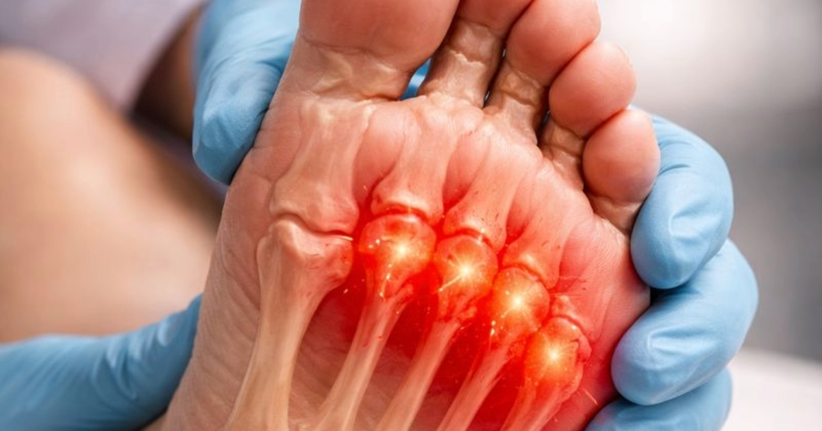
The Great Fat Pad Migration of Metarsalgia
4 January 2026
By Mark Reyneker, B.T. Pod (SA), MSc (SA)
The plantar fat pad is a specialized collection of adipocytes (fat cells) encased in a "regular arrangement" of fibrous tissue septa. These septa are rich in elastin to provide mechanical strength and are anchored firmly to both the skin and the proximal phalanges (toe bones).
This architecture makes the fat pad a "prototypical shock absorber," with the density of sensory nerves at the metatarsal heads (MTHs) being twice as high as in proximal regions to help the body detect and manage weight-bearing stimuli.
The Evolution of Pathology: Atrophy vs. Migration
Historically, researchers believed that fat pads in the foot simply atrophied (shrank) due to diabetes.
• The Atrophy Theory: Suggested that high blood sugar (glycation) caused collagen fibrils to become distorted and fragmented, leading to a 30% reduction in surface area and a 16% decrease in the diameter of fat cells in the heel.
• The Migration Theory: Newer research indicates that in the forefoot, the number and size of fat cells often remain normal, but they shift position. Studies found no significant difference in fat cell size between diabetic and healthy individuals, suggesting the "thinning" felt by patients is actually a mechanical displacement.
Quantifying the Distal Shift
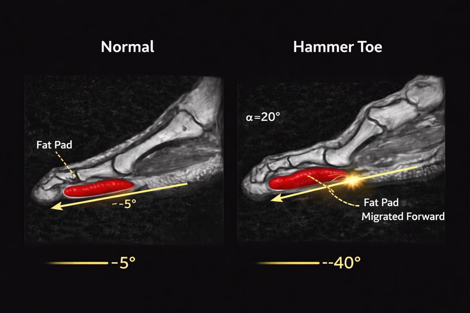
When a hammer or claw toe deformity occurs, the metatarsophalangeal (MTP) joint hyperextends. Because the fat pad is tethered to the toe bone, this upward bend creates a "distal pull," dragging the cushion forward.
The sources provide precise data on this shift:
• Sub-MTH Thinning: In feet with toe deformities, the protective fat under the metatarsal head drops from an average of 6.0 mm to just 2.5 mm.
• Sub-phalangeal Thickening: As the fat is pulled forward, the padding under the toe bone increases from 7.6 mm to 9.1 mm.
• The 65% Reduction: The "thickness ratio" (the balance of fat between the ball of the foot and the toe) decreases by 65% in deformed feet.
• Direct Correlation: There is a powerful 0.85 correlation between the severity of the toe angle and the displacement of the fat pad—meaning the more the toe "claws," the further the cushion migrates away from the bone.
Structural Breakdown: Rupture and Exposure
In the most severe cases of deformity, the tissue does more than just move; it breaks. In nearly one-half (50%) of deformed toes studied via MRI, the fat tissue was found to be discontinuous or almost completely absent under the metatarsal head. This suggests that the distal pull can become so extreme that the plantar fat pad actually ruptures or separates, leaving the MTH "prominent and unprotected" against the ground.
Clinical Consequences and New Frontiers
This migration transforms the ball of the foot into a high-pressure zone. This structural shift is a significant predictor of elevated plantar pressure, which is the leading risk factor for skin trauma and ulceration.
Understanding this process and detecting it early is what we specialize in at the Family Podiatry Centre. We use EMED scanning equipment and gait analyses to trace changes that are occurring in the forefoot metatarsal area. For more on treatment read "Treating Metatarsalgia Caused By Fat Pad Pathology".
Understanding this process and detecting it early is what we specialize in at the Family Podiatry Centre. We use EMED scanning equipment and gait analyses to trace changes that are occurring in the forefoot metatarsal area. For more on treatment read "Treating Metatarsalgia Caused By Fat Pad Pathology".
Book An Appointment
Disclaimer: The word "treatment" in this article refers to the care and management of a patient’s health to prevent, cure, or improve a condition. Treatment results vary and do not necessarily indicate a cure. This article is for informational and educational purposes only and does not constitute medical advice.
About the author
Mark Reyneker is a podiatrist and human gait specialist with 8 years of training and over 25 years of clinical experience. He is the Founder and Clinical Director of Family Podiatry Centre and has a Bachelors degree in Podiatric Medicine and a Master’s degree in paleoanthropology, with research focused on human foot function and metatarsal loading.