Fascia: What It Is, What It Does and Why It Hurts



18 September 2026
By Mark Reyneker, BTech (Podiatry), MSc (Palaeontology), Registered Podiatrist, Foot & Gait Specialist | Founder & Clinical Director, Family Podiatry Centre
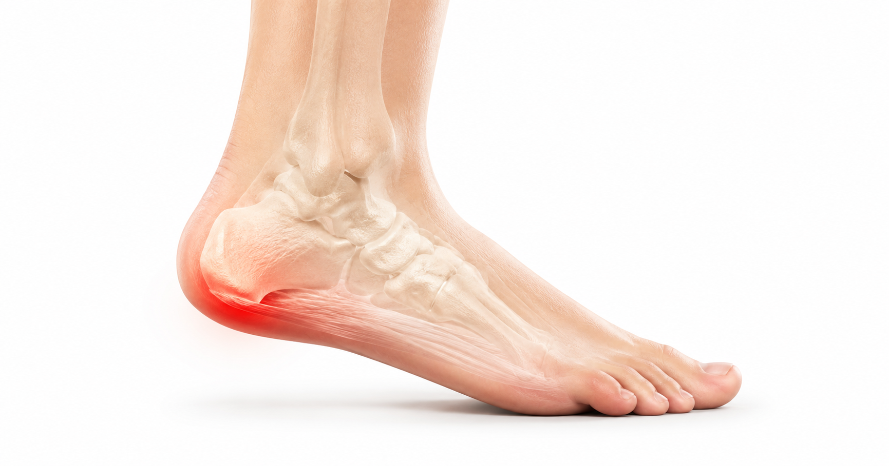
Fascia is a living network of collagen-rich connective tissue that surrounds, separates and connects muscles, bones, nerves and other structures throughout the body. It helps transmit force, supports movement, allows tissues to glide against each other and contains sensory nerve endings that can contribute to pain and body awareness.
Fascia: What It Is, What It Does and Why It Can Hurt
That view has changed considerably.
Fascia is now understood as a biologically active, mechanically responsive and richly innervated connective tissue. It helps transmit force, allows neighbouring structures to slide relative to each other, contributes to proprioception, responds to mechanical loading and can itself become a source of pain. Even the definition of fascia continues to evolve.
A major 2025 anatomical paper proposed viewing fascia as a layered body-wide connective-tissue network containing relatively stiff collagenous layers separated by more mobile, hydrated interfaces. Other anatomists have argued that describing fascia as an anatomical “system” goes too far. The terminology is still being refined, but the underlying biology is becoming much clearer (Stecco et al., 2025).
What exactly is fascia?
The crural fascia surrounding the muscles of the lower leg, for example, has been shown histologically to contain approximately three layers of collagen bundles separated by very thin layers of loose connective tissue. The mean thickness in one anatomical study was approximately 0.9 mm, although fascial thickness varies considerably according to anatomical location (Stecco et al., 2009).
Fascia therefore has two apparently contradictory requirements. It must be strong enough to transmit tension. At the same time, its layers must be capable of sliding. Much of fascial biology appears to revolve around balancing these two properties.
What is fascia actually made from?
Type I collagen provides enormous tensile strength.
Type VI collagen helps organise the extracellular matrix around cells and may influence the mechanical environment in which fascial cells live.
This is one of the areas in which fascia research is moving particularly quickly. The fascia is therefore not simply a sheet of collagen. It is an organised extracellular matrix in which different collagen molecules, cells, water and ground substance interact.
Collagen crimp: nature’s mechanical reserve
During the early part of loading, some of this crimp straightens. As loading continues, progressively more collagen fibres become engaged. This contributes to fascia’s characteristic non-linear mechanical behaviour: the tissue initially deforms relatively easily and then becomes progressively harder to stretch (Stecco et al., 2009). This behaviour is particularly relevant to the foot, where connective tissues repeatedly move between relatively compliant and relatively stiff states during every step.
Fascia contains relatively little elastin
Much of fascia’s ability to lengthen and recoil therefore arises from collagen crimp, fibre recruitment, fluid movement and the interaction between its layers rather than simply from large quantities of elastin. Fascia behaves viscoelastically, rather than behaving like a rubber band.That difference matters. A viscoelastic tissue responds differently depending upon the magnitude, speed and duration of the load applied to it.
The forgotten part of fascia: water and ground substance
Human studies have demonstrated that the amount of hyaluronan differs substantially between different fasciae depending upon their mechanical requirements. Structures requiring considerable sliding, such as retinacula around joints, can contain substantially more hyaluronan than fascia tightly adherent to underlying muscle (Fede et al., 2018). This gives us another way of thinking about fascia. There is the load-bearing collagen component, and there is the sliding interface between the collagen layers. Both matter.
Fasciacytes: cells that appear to regulate fascial gliding
Fascia is not simply collagen manufactured at some point during development and subsequently left in the body. Its extracellular environment is continually being maintained and modified by living cells. Alongside fasciacytes are conventional fibroblasts, vascular cells, immune cells and, particularly in injured or fibrotic tissue, myofibroblasts.
Mechanical load can influence these cells. This means that fascia is capable of remodelling in response to the forces placed upon it.
Fascia is also a sensory tissue
A systematic review of fascial innervation identified free nerve endings, proprioceptive structures and nerve fibres associated with blood vessels and collagen bundles. Both nociceptive fibres—those capable of signalling potentially damaging stimuli—and mechanosensitive receptors have been demonstrated (Suarez-Rodriguez et al., 2022). That immediately changes the discussion around fascial pain.
Pain attributed to fascia does not necessarily have to originate in the muscle sitting underneath it. The fascia itself can generate sensory information.
Human experimental studies have produced similar findings elsewhere in the body. When irritating solutions have been injected specifically into deep fascia, they can produce intense and sometimes more prolonged pain than equivalent stimulation of nearby muscle (Schilder et al., 2014). Another experiment using nerve growth factor demonstrated that fascial nociceptors could remain sensitised to mechanical and chemical stimulation for days after the original exposure (Deising et al., 2012).
This introduces an important concept. Fascial pain does not require the fascia to be torn. A mechanically intact fascia can potentially become sensitised.
Fascia also contributes to proprioception
Fascia appears to be one component of that sensory network. This is especially plausible in the legs, where large fascial structures cross joints, receive muscular attachments and change tension continuously during walking and running.
The fascia of the leg
The fascia lata is now recognised as a regionally specialised, multilayered collagen structure receiving contributions from muscles including gluteus maximus and tensor fasciae latae. Its architecture allows it to participate in force transmission and lower-limb stability rather than functioning purely as a passive wrapper (Stecco et al., 2026).
The crural fascia: the sleeve around the lower leg
The iliotibial band and fascial pain
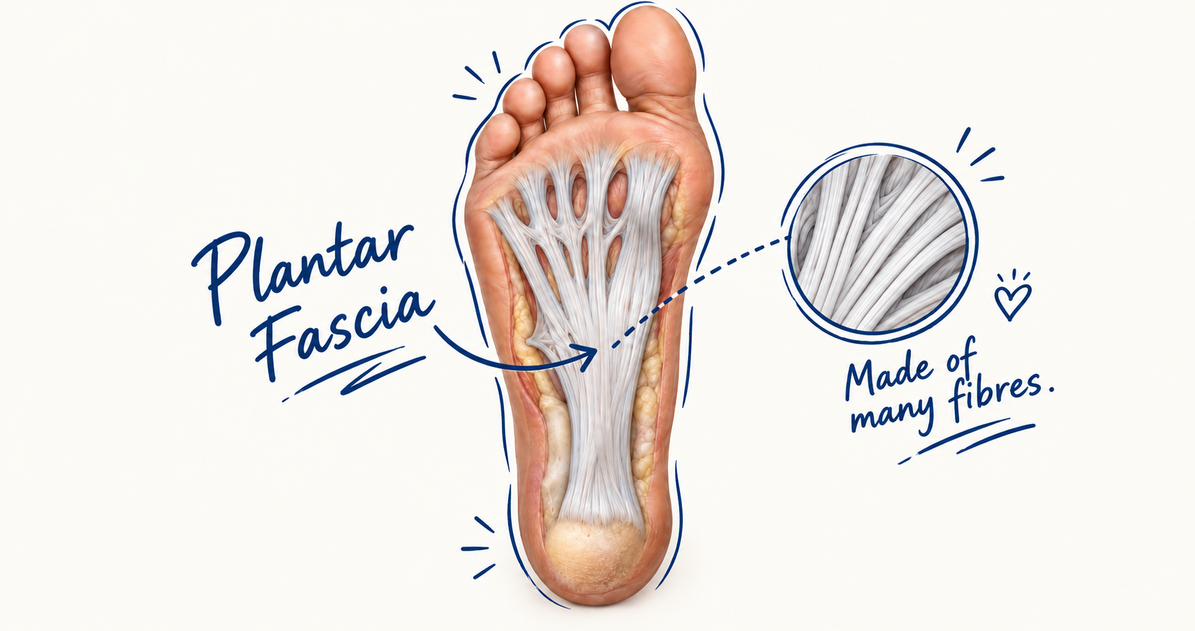
And then there is the plantar fascia
Describing it literally as the thickest fascia in the human body is more difficult because fascial thickness varies dramatically by location, individual and measurement technique. What is clear is that the central plantar aponeurosis is exceptionally robust for its size and has an unusually important mechanical job.
The plantar fascia is built in the direction it is loaded
What does the plantar fascia actually do?
The windlass mechanism
Under single-leg loading, intrinsic muscle stiffness increased dramatically, showing that arch control is not simply a passive fascial event (Shinohara et al., 2025). The foot is therefore neither purely passive nor purely muscular. It is a muscle–fascia–ligament–bone system in which several structures share load.
Fascia as a spring
Running—particularly non-rearfoot running—places greater emphasis on spring-like behaviour than ordinary walking (Davis and Challis, 2023). Once again, the plantar fascia is only part of this system. Intrinsic muscles and other passive structures also store, absorb or generate energy.
The Achilles tendon and plantar fascia
When healthy fascia becomes painful
Fasciopathy rather than simply fasciitis
Yet histological examination of chronic plantar fascia specimens has frequently found collagen degeneration, disorganisation and myxoid change rather than the cellular picture expected from a predominantly inflammatory disorder. This led Lemont and colleagues to propose the term plantar fasciosis more than two decades ago (Lemont et al., 2003).
More recent literature generally supports the idea that chronic plantar fasciitis is substantially a degenerative/remodelling disorder, although this does not mean inflammatory signalling is completely absent, particularly earlier in the disease process (Tseng et al., 2023). For that reason, plantar fasciopathy is often the most biologically neutral description. “Plantar fasciitis” remains the term most patients and clinicians recognise.
What changes inside a painful plantar fascia?
Thickness greater than approximately 4 mm near the calcaneal origin is commonly used as an imaging feature supporting plantar fasciopathy. It is not, however, a perfect dividing line between healthy and painful tissue. Structural change and pain are related, but they are not identical.
Is a painful plantar fascia softer or stiffer?
The enthesis matters
This may help explain why pathology repeatedly develops at or close to the calcaneal attachment rather than randomly along the fascia. It also explains why a simple model of the plantar fascia being “pulled too hard” does not capture the entire problem.
Body mass and plantar fascial load
Greater body mass changes repetitive load, plantar pressure, muscular demand and tissue deformation. Metabolic factors associated with obesity may also influence connective-tissue biology.
Age, diabetes and collagen
With ageing—and particularly with chronic hyperglycaemia—these cross-links can alter the mechanical behaviour and turnover of collagen-rich connective tissues (Paul and Bailey, 1996). This provides a plausible biological connection between ageing, diabetes and altered mechanics within tendons, ligaments and fascia. Diabetes is also associated clinically with plantar fascial abnormalities and plantar heel pain (Gariani et al., 2020).
Fascial gliding and “densification”
The clinical science is less mature than the basic science, however. We do not yet have a universally accepted clinical test that can reliably identify a painful fascial “densification”, nor can every sensation of tightness reasonably be attributed to altered hyaluronan. This remains an active area of research rather than a complete explanation for myofascial pain.
Fibrosis is different from densification
Plantar fibromatosis: when the fascia actually grows nodules
Histologically the disease involves proliferating fibroblasts and myofibroblasts with abnormal collagen deposition. It belongs to the same broad family of fibroproliferative disorders as Dupuytren disease of the hand (Tersago and Constantin, 2025). This is genuine structural disease of the plantar fascia rather than the overload-associated fasciopathy responsible for most plantar heel pain.
Fascial strains and tears
Ultrasound or MRI may demonstrate disruption of the normally continuous fascial fibres. Acute rupture is uncommon compared with plantar fasciopathy but is a well-recognised plantar-fascial injury (Theodorou et al., 2000).
Pain does not always match structural damage
Pathological fascia has been associated with changes in nociceptive nerve density, inflammatory signalling and nociceptor sensitisation (Kondrup et al., 2022). Once nociceptors become sensitised, forces that were previously tolerated may generate a stronger pain response. Persistent nociceptive input may subsequently influence processing higher within the nervous system. Pain therefore becomes more complicated than simply asking: How damaged is the collagen? The state of the sensory system matters as well.
Not every pain beneath the heel comes from fascia
Current research suggests it may account for a meaningful proportion of cases traditionally grouped under nonspecific heel pain (Yi et al., 2022). Nerve irritation—including branches of the lateral plantar nerve—can mimic plantar fascial pain. Calcaneal stress injury can do the same. So can inflammatory arthropathies and other enthesopathies. This is one reason why imaging a thick plantar fascia does not automatically establish that the fascia is the sole pain generator.
The same applies to shin pain
A 2025 scoping review found continuing interest in traction theories but also substantial evidence supporting multiple biomechanical and loading-related contributors (Almubarak et al., 2025). The anatomy overlaps. The pathology does not always belong to a single tissue.
Why fascia hurts: a broader model
Pressure inside a fascial compartment can rise. Nociceptors within the fascia can become sensitised. Inflammatory and neuroimmune signalling can amplify those responses. And neighbouring nerves, muscle, fat or bone may contribute to a painful region that is clinically described as “fascial”. That complexity is probably closer to reality than the older idea that painful fascia is simply “tight”.
Fascia is neither passive wrapping nor a magical body-wide web
- It has cells.
- It has blood vessels.
- It contains extracellular matrix.
- It contains nerves.
- It responds to mechanical loading.
- It transmits force.
- Its layers glide against one another.
- It helps organise muscle compartments.
- It interacts mechanically with muscles and bones.
And under certain circumstances it can become an important source of pain.
The amount of force transmitted over very long proposed “myofascial chains”, however, and the clinical importance of many remote fascial connections remain much less certain than the anatomical continuity itself. Local and regional force transmission are well supported. Very large claims about whole-body fascial chains deserve more cautious interpretation.
The plantar fascia may be the clearest example of what fascia really is
Perhaps most importantly, modern fascia research is gradually moving us away from thinking about tissues in isolation. The foot does not operate as bones plus muscles plus tendons plus ligaments plus fascia. These structures continually load one another. The plantar fascia is part of that mechanical conversation.
Every step stretches it, unloads it, changes its tension, alters its relationship with the intrinsic muscles and modifies the shape of the arch. For most of our lives it manages this remarkably well. Plantar fasciopathy appears when the balance between mechanical demand, tissue capacity, biological repair and sensory processing begins to shift.
That may be a considerably more useful way of understanding plantar fascial pain than imagining a simple inflamed band beneath the heel.
Book An Appointment
Disclaimer: The word "treatment" in this article refers to the care and management of a patient’s health to prevent, cure, or improve a condition. Treatment results vary and do not necessarily indicate a cure. This article is for informational and educational purposes only and does not constitute medical advice.
About the Author
Mark B. Reyneker, BTech (Podiatry), MSc (Palaeontology) is a podiatrist and Founder & Clinical Director of Family Podiatry Centre, with more than 25 years of clinical experience across South Africa, Malaysia and Singapore. His clinical interests include foot and lower-limb pain, gait and biomechanics, sports-related foot conditions, orthotic therapy and footwear.
Alongside his clinical practice, Mark conducts research into human gait and foot biomechanics. His MSc research at the University of the Witwatersrand investigated human propulsion and the structural properties of the metatarsals. He is also the inventor of A Foot Orthotic, an orthotic technology developed through an international patent family.
References
- Adstrum, S., Hedley, G., Schleip, R., Stecco, C. and Yucesoy, C.A. (2017) ‘Defining the fascial system’, Journal of Bodywork and Movement Therapies, 21(1), pp. 173–177.
Original article:(PubMed) - Albano, D., Messina, C., Gitto, S. et al. (2023) ‘Shear-wave elastography of the plantar fascia: a systematic review and meta-analysis’, Journal of Ultrasound, 26, pp. 59–64.
Original article: (Springer) - Almubarak, A. et al. (2025) ‘Medial Tibial Stress Syndrome: A Scoping Review of Epidemiology, Biomechanics, and Risk Factors’.
Original article: (PubMed) - Chen, D.W. et al. (2014) ‘Anatomy and biomechanical properties of the plantar aponeurosis: a cadaveric study’.
Original article: (PubMed) - Cheung, J.T.M., Zhang, M. and An, K.N. (2006) ‘Effect of Achilles tendon loading on plantar fascia tension in the standing foot’, Clinical Biomechanics.
Original article:(PubMed) - Davis, D.J. and Challis, J.H. (2023) ‘Characterizing the mechanical function of the foot’s arch across steady-state gait modes’, Journal of Biomechanics, 151, 111529.
Original article: (PubMed) - Deising, S., Weinkauf, B., Blunk, J., Obreja, O., Schmelz, M. and Rukwied, R. (2012) ‘NGF-evoked sensitization of muscle fascia nociceptors in humans’, Pain, 153, pp. 1673–1679.
Original article: (PubMed Central (PMC)) - Fede, C., Angelini, A., Stern, R. et al. (2018) ‘Quantification of hyaluronan in human fasciae: variations with function and anatomical site’, Journal of Anatomy, 233(4), pp. 552–556.
Original article: (PubMed) - Fede, C., Clair, C., Petrelli, L. et al. (2026) ‘Redefining the Collagen Composition of Human Fasciae: Emerging Collagen Types and Structural Heterogeneity’, International Journal of Molecular Sciences, 27(2), 1021.
Original article: (PubMed) - Gariani, K. et al. (2020) ‘Plantar Fasciitis in Diabetic Foot Patients: Risk Factors, Pathophysiology, Diagnosis, and Management’.
Original article: (PubMed) - Hamstra-Wright, K.L. et al. (2021) ‘Risk Factors for Plantar Fasciitis in Physically Active Individuals: A Systematic Review and Meta-analysis’.
Original article: (PubMed) - Kondrup, F., Gaudreault, N. and Venne, G. (2022) ‘The deep fascia and its role in chronic pain and pathological conditions: A review’, Clinical Anatomy, 35(5), pp. 649–659.
Original article: (PubMed) - Lemont, H., Ammirati, K.M. and Usen, N. (2003) ‘Plantar fasciitis: a degenerative process (fasciosis) without inflammation’.
Original article: (PubMed) - Ortiz-Miguel, S., Miguel-Pérez, M., Blasi, J. et al. (2023) ‘Compartments of the crural fascia: clinically relevant ultrasound, anatomical and histological findings’, Surgical and Radiologic Anatomy, 45, pp. 1603–1617.
Original article: (DOI) - Paul, R.G. and Bailey, A.J. (1996) ‘Glycation of collagen: the basis of its central role in the late complications of ageing and diabetes’, International Journal of Biochemistry & Cell Biology, 28(12), pp. 1297–1310.
Original article: (PubMed) - Pettenuzzo, S., Terzano, M., Todesco, M. et al. (2026) ‘Histological analysis, viscoelastic characterization, and modeling of human plantar fascia’, Acta Biomaterialia, 217, pp. 488–502.
Original article: (PubMed) - Schilder, A., Hoheisel, U., Magerl, W. et al. (2014) ‘Sensory findings after stimulation of the thoracolumbar fascia with hypertonic saline suggest its contribution to low back pain’, Pain.
Original article: (PubMed) - Shinohara, H., Umezaki, Y., Ikeda, A. et al. (2025) ‘Reconsideration of the load-bearing functions of the plantar fascia and intrinsic foot muscles in the windlass mechanism’, Scientific Reports, 15, 12923.
Original article: (PubMed) - Stecco, C., Pavan, P.G., Porzionato, A. et al. (2009) ‘Mechanics of crural fascia: from anatomy to constitutive modelling’, Surgical and Radiologic Anatomy, 31(7), pp. 523–529.
Original article: (PubMed) - Stecco, C., Stern, R., Porzionato, A. et al. (2011) ‘Hyaluronan within fascia in the etiology of myofascial pain’, Surgical and Radiologic Anatomy.
Original article: (PubMed) - Stecco, C., Fede, C., Macchi, V. et al. (2018) ‘The fasciacytes: A new cell devoted to fascial gliding regulation’, Clinical Anatomy, 31(5), pp. 667–676.
Original article: (PubMed) - Stecco, C., Pratt, R., Nemetz, L.D., Schleip, R., Stecco, A. and Theise, N.D. (2025) ‘Towards a comprehensive definition of the human fascial system’, Journal of Anatomy, 246(6), pp. 1084–1098.
Original article: (PubMed) - Suarez-Rodriguez, V., Fede, C., Pirri, C. et al. (2022) ‘Fascial Innervation: A Systematic Review of the Literature’, International Journal of Molecular Sciences, 23(10), 5674.
Original article: (PubMed) - Taguchi, T., Yasui, M., Kubo, A. et al. (2013) ‘Nociception originating from the crural fascia in rats’, Pain, 154(7), pp. 1103–1114.
Original article: (PubMed) - Thanwisate, T., Siriwanarangsun, P., Piyaselakul, S., Tharmviboonsri, T. and Chuckpaiwong, B. (2024) ‘Plantar Fascia Thickness and Stiffness in Healthy Individuals vs Patients With Plantar Fasciitis’, Foot & Ankle International, 45(11), pp. 1270–1278.
Original article: (PubMed) - Tersago, J. and Constantin, A. (2025) ‘Soleful solutions: Advancements in treatment strategies for Ledderhose disease’, Foot and Ankle Surgery, 31(1), pp. 10–14.
Original article: (PubMed) - Tseng, W-C., Chen, Y-C., Lee, T-M. and Chen, W-S. (2023) ‘Plantar Fasciitis: An Updated Review’, Journal of Medical Ultrasound, 31(4), pp. 268–274.
Original article: (PubMed Central (PMC))
Frequently Asked Questions
Yes. Fascia is not simply an inert wrapping around muscles. Deep fascia contains sensory nerve fibres, including nociceptive fibres capable of detecting potentially damaging mechanical and chemical stimuli. Experimental studies have shown that stimulation of fascia can produce significant pain, and in some experiments fascial stimulation produced more prolonged pain than stimulation of the underlying muscle. This means that pain felt in the calf, thigh or foot can potentially arise partly from the fascia itself rather than always originating from muscle, tendon or bone. The situation becomes more complex in persistent pain because nociceptors within fascia can become sensitised. Once sensitised, relatively normal mechanical forces may produce a disproportionately strong pain response. Fascia should therefore be considered as one potential pain-generating tissue within the wider musculoskeletal system rather than the explanation for every unexplained muscular pain (Weiss and Kalichman, 2021).
There is no single sensation that identifies fascial pain. People may describe aching, pulling, pressure, tenderness, tightness or a deeper poorly localised discomfort. Pain may become more noticeable when the tissue is stretched, compressed or loaded. Experimental research suggests that deep fascia can generate relatively diffuse pain because it contains nociceptive nerve endings capable of responding to mechanical, chemical and inflammatory stimuli (Weiss and Kalichman, 2021). In the foot, plantar fascial pain tends to be much easier to localise. It commonly occurs around the medial plantar heel near the calcaneal attachment of the plantar fascia and may extend forwards into the arch. Burning, tingling, electric-shock sensations or numbness raise the possibility that a nerve is contributing to the symptoms. Neural causes of plantar heel pain include entrapment of branches of the tibial or lateral plantar nerves and can sometimes resemble plantar fasciopathy (Alshami, Souvlis and Coppieters, 2008).
First-step pain after getting out of bed—or pain when standing after sitting for a long period—is one of the classic clinical features of plantar fasciopathy. During prolonged rest, the plantar fascia and surrounding tissues are relatively unloaded. When the foot suddenly accepts bodyweight again, the arch deforms and the plantar fascia is rapidly tensioned. A sensitised or structurally altered plantar fascia may respond strongly to this sudden return of load. Interestingly, the pain often improves after several minutes of walking as the tissues begin moving and loading more continuously. It may then return later after prolonged standing, walking or running. The precise biological explanation for first-step pain has not been completely established, so it is better regarded as a characteristic clinical behaviour of plantar fasciopathy rather than proof of a particular microscopic mechanism (May and Judy, 2002). Persistent morning heel pain can also occur with other conditions, so the symptom alone does not confirm that the plantar fascia is responsible.
Sometimes, but the term plantar fasciitis probably oversimplifies what happens in many chronic cases. The suffix -itis implies inflammation. Histological studies of chronically painful plantar fascia, however, have commonly demonstrated collagen degeneration, disorganisation and changes within the extracellular matrix rather than the cellular appearance of a purely inflammatory disorder. This led to the term plantar fasciosis, while many researchers and clinicians now prefer plantar fasciopathy because it does not assume that inflammation or degeneration is the only process involved. Inflammatory signalling may still occur, particularly during periods of acute irritation or earlier disease. The modern view is therefore not that inflammation is completely absent, but that long-standing plantar heel pain cannot usually be explained simply as an inflamed fascia. That distinction may also explain why chronic plantar fascia pain can persist long after the original increase in activity or mechanical loading has passed. Lemont, Ammirati and Usen (2003) famously described chronic plantar fasciitis as a degenerative process rather than conventional inflammation.
They can. The gastrocnemius–soleus complex and Achilles tendon influence how the ankle and foot move during walking and running. Limited ankle dorsiflexion can alter the way the body progresses over the foot and potentially increase loading through the plantar structures. There is also a mechanical relationship between Achilles tendon tension, the calcaneus and plantar fascia. This does not mean that every person with plantar fasciopathy simply has “tight calves”, but restricted ankle movement and gastrocnemius tightness can be relevant in some patients. This relationship is strong enough that gastrocnemius recession has been studied as a treatment for severe, persistent plantar fasciopathy associated with gastrocnemius contracture. A recent systematic review of randomised trials continued to support a biomechanical relationship between gastrocnemius restriction and increased plantar fascial tension (Arshad et al., 2026). Calf function therefore forms part of the assessment, alongside foot mechanics, activity levels, footwear, bodyweight, strength and the location of the pain itself.
One possible explanation is chronic exertional compartment syndrome, although there are many other causes of exercise-related lower-leg pain. The muscles of the lower leg sit inside compartments surrounded by relatively strong fascia. During exercise, muscle volume and local pressure increase. In chronic exertional compartment syndrome, pressure within one or more compartments rises sufficiently to produce pain, pressure, tightness and sometimes neurological symptoms. A characteristic pattern is symptoms appearing after a relatively predictable period or intensity of exercise and improving after activity stops. This differs considerably from ordinary post-exercise muscle soreness. Chronic exertional compartment syndrome is also an excellent example of how fascia can contribute to pain without the fascia itself necessarily being injured. The relatively non-compliant fascial compartment influences the mechanical environment surrounding the muscles, blood vessels and nerves (Vajapey and Miller, 2017). Other causes—including tibial bone stress, medial tibial stress syndrome, nerve irritation and muscular injury—need to be considered in runners with recurrent lower-leg pain.
Yes, although plantar fascia rupture is much less common than plantar fasciopathy. A tear may occur suddenly during running, jumping or another high-load activity. Some people describe a sudden sharp pain or a sensation of something giving way beneath the foot. Swelling, bruising and difficulty walking may follow. Plantar fascia rupture can occur in an otherwise healthy athlete, but the literature suggests that it occurs more frequently in people who already have plantar fasciopathy. Corticosteroid injection has also been associated with plantar fascia rupture. A systematic review involving 157 affected feet found that the majority of reported patients had pre-existing plantar fasciitis and many had previously received local corticosteroid injections. Most reported cases were managed conservatively, although the available evidence consisted predominantly of case series rather than high-quality randomised trials (Debus et al., 2021). A sudden change from chronic heel pain to severe acute pain deserves reassessment rather than being assumed to be another flare of plantar fasciitis.
Imaging can show structural changes within the plantar fascia, but an image does not perfectly measure pain. Ultrasound may demonstrate increased plantar fascia thickness, reduced echogenicity, disruption of the normal fibre pattern, increased vascularity or a tear. MRI can demonstrate plantar fascia thickening, altered signal, surrounding soft-tissue oedema and abnormalities within the calcaneus. A systematic review and meta-analysis of 42 studies involving 2,928 participants found that people with plantar heel pain were more likely to demonstrate plantar fascia thickening and other structural abnormalities on ultrasound and MRI than people without heel pain (Drake et al., 2022). However, structural abnormalities can remain after the pain has disappeared. Long-term research has shown that some symptom-free people continue to have an abnormal-looking plantar fascia years later. Imaging is therefore most useful when interpreted alongside the history and clinical examination rather than being considered a direct pain detector (Hansen et al., 2018).
They can help some people, although the explanation is probably more complicated than physically “breaking up” or permanently lengthening fascia. Plantar fascia-specific stretching has been investigated in several clinical trials. A systematic review found some evidence that plantar fascia-specific stretching can reduce pain, although the overall quality of the research varied considerably (Siriphorn and Eksakulkla, 2020). A very recent systematic review published in 2026 found that stretching was not clearly superior to other conservative interventions overall, highlighting how variable individual responses and treatment protocols can be. Manual myofascial techniques have also produced improvements in plantar heel pain in clinical trials. Ajimsha, Binsu and Chithra (2014), for example, reported reductions in pain and disability following myofascial release compared with a control intervention. These treatments may influence pain sensitivity, tissue movement, joint mobility and neuromuscular function. Evidence that massage physically “breaks apart adhesions” within normal fascia is considerably less convincing.
Plantar fasciopathy can improve within weeks or months, but some cases persist much longer. A recent study following 200 patients over one year found progressive improvements in pain, function and quality of life, with the largest improvement occurring during the first three months (Riel et al., 2025). Longer-term research shows how variable the condition can be. Hansen et al. followed patients for an average of almost ten years and found that the average duration of symptoms among those who eventually became asymptomatic was approximately two years. A subgroup continued to experience symptoms many years after the original diagnosis. Interestingly, plantar fascia thickness did not reliably predict who remained painful (Hansen et al., 2018). This reinforces an important feature of fasciopathy: recovery is not simply a matter of waiting for damaged collagen to “heal”. Mechanical loading, tissue capacity, strength, bodyweight, sensitisation, activity and the accuracy of the original diagnosis can all influence the course of symptoms. Persistent heel pain also deserves reassessment because not every painful heel is plantar fasciopathy. Heel fat-pad disorders, nerve entrapment, calcaneal stress injury and other conditions may produce similar symptoms (Chang et al., 2022).