Why the Big Toe Matters: How Evolution Shaped the Human Foot for Walking and Running



21 September 2026
By Mark Reyneker, BTech (Podiatry), MSc (Palaeontology), Registered Podiatrist, Foot & Gait Specialist | Founder & Clinical Director, Family Podiatry Centre
Why is the big toe so important?
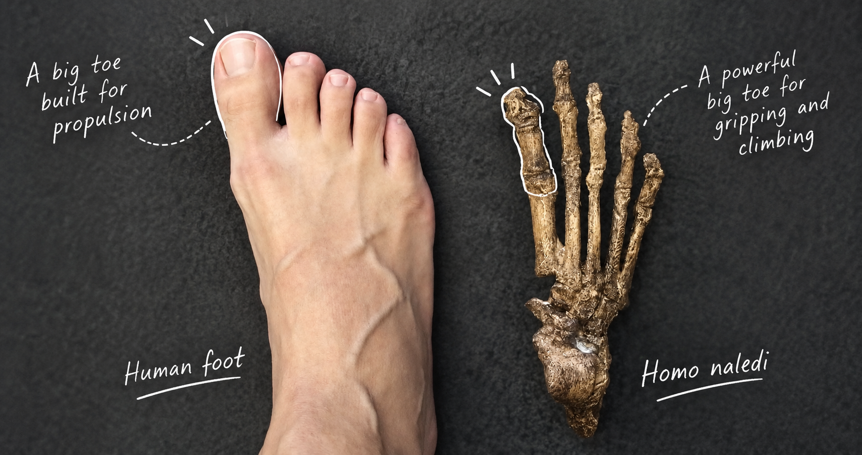
Its importance also reflects our evolutionary history. Early primate-like ancestors had a more divergent big toe suited to grasping. During human evolution, the hallux became aligned with the other toes as the foot became increasingly adapted for habitual walking on two legs.
The result is the distinctive human big toe: a large, relatively straight digit integrated with the first metatarsal, sesamoids, plantar fascia and foot muscles. Pain, arthritis, deformity or loss of movement at this joint can therefore change how forces pass through the foot during walking and running.
The Big Toe: How Evolution Turned a Grasping Digit into the Engine of the Human Foot
As a podiatrist, however, I spend a considerable amount of time thinking about the big toe. Pain, stiffness, arthritis, hallux valgus, sesamoid problems and restricted movement of the first metatarsophalangeal joint can all alter the way a person walks. Sometimes the change is subtle. Sometimes the entire propulsive strategy of the foot appears to change.
Because the human big toe is not simply a large version of our other toes. It represents one of the most important transformations in the evolution of the human foot.
Our distant primate ancestors possessed feet capable of grasping. The first digit could diverge from the other toes and participate in gripping branches. During hominin evolution, that arrangement changed dramatically. The hallux moved into line with the other toes, became incorporated into a progressively stiffer forefoot and eventually became an important component of the propulsive system used during human walking and running. In other words, evolution largely sacrificed prehension for propulsion. And the anatomy of your big toe today still carries the evidence of that transformation.
The big toe is really a mechanical system
The functional unit includes the:
- first metatarsal
- proximal and distal hallucal phalanges
- first metatarsophalangeal joint
- first tarsometatarsal joint
- medial cuneiform
- tibial and fibular sesamoids
- plantar plate and joint capsule
- plantar fascia
- flexor hallucis longus
- flexor hallucis brevis
- extensor hallucis longus
- abductor hallucis
- adductor hallucis
- and several ligamentous structures.
The first metatarsal is substantially more robust than the lesser metatarsals. Beneath its head sit two sesamoid bones embedded within the tendons of flexor hallucis brevis. These sesamoids are not insignificant accessory bones. They increase the mechanical advantage of the flexor apparatus, help transmit considerable loads beneath the first metatarsal head and provide a sophisticated pulley system around which the first metatarsophalangeal joint operates. This is already a clue to the importance of the medial forefoot. The human foot has effectively constructed a reinforced mechanical apparatus beneath the big toe.
Before propulsion came grasping
In modern humans, the hallux is strongly adducted — positioned approximately parallel with the remaining toes. In African apes it is considerably more abducted, allowing the foot to participate in grasping.That distinction reflects fundamentally different locomotor requirements.
Ardipithecus: a big toe that still grasped
Lovejoy and colleagues (2009) interpreted the Ardipithecus foot as a fascinating locomotor compromise: capable of terrestrial bipedalism but retaining a powerful hallucal grasping mechanism useful in an arboreal environment. Whatever the exact reconstruction of Ardipithecus locomotion — an area that remains debated — one point is particularly important.The modern human propulsive hallux had not yet appeared. The big toe was still, to a considerable degree, a grasping digit.
Something remarkable had happened by 3.66 million years ago
- an adducted hallux
- a functional longitudinal arch
- lateral-to-medial transfer of loading
- and propulsion involving the hallux.
Indeed, comparative studies of metatarsophalangeal morphology suggest that A. afarensis probably differed from modern humans in forefoot dorsiflexion and push-off mechanics (Fernández et al., 2016). But something fundamental had changed. The big toe was becoming integrated into the direction of travel. The foot was becoming a propulsive structure.
The rest of the foot was changing too
The human lineage developed a suite of interacting features including:
- hallucal adduction
- shorter toes
- changes in metatarsal head morphology
- increasing midfoot stiffness
- longitudinal arch development
- transverse arch development
- changes in plantar soft tissues
- altered muscular function
- and changes in the geometry and loading of the metatarsals.
Venkadesan and colleagues (2020) demonstrated that the transverse arch contributes substantially to longitudinal foot stiffness. This matters enormously when thinking about evolution. A propulsive hallux is useful only if the structures behind it can transmit force effectively. The evolution of the big toe therefore occurred as part of the evolution of an integrated mechanical system.
From a grasping foot to a spring-loaded lever
Now the foot must provide sufficient stiffness to transmit force. The human foot therefore behaves less like a simple rigid lever and more like a dynamically adjustable mechanical structure. This is where the hallux becomes particularly important. As the heel rises during late stance, the first metatarsophalangeal joint dorsiflexes. That apparently simple movement influences structures extending far beyond the toe itself.
The big toe and the windlass mechanism
In the foot, dorsiflexion of the hallux increases tension within the plantar aponeurosis and can contribute to elevation and stiffening of the longitudinal arch. This is the famous windlass mechanism. The concept remains extremely influential in podiatry.
Modern biomechanics, however, suggests that the foot is more complicated than a purely passive windlass. The plantar fascia can store and return elastic energy, the intrinsic muscles actively influence foot behaviour, and both longitudinal and transverse architecture contribute to stiffness. The windlass is therefore best understood as one component within a much larger integrated system rather than as the single explanation for human foot function.
The plantar fascia may itself have an evolutionary history
Farris and colleagues (2020) proposed that hominins may subsequently have evolved a thicker and stiffer plantar aponeurosis as the longitudinal arch evolved, enhancing both windlass function and elastic energy storage. This is a beautiful example of evolutionary modification. Evolution does not always invent an entirely new structure. Sometimes an existing structure acquires a new mechanical role.
Why the human big toe bends upwards
This relates partly to the shape of our metatarsal heads. As the heel rises, the leg and body continue moving forward while the hallux remains in contact with the ground. The first MTP joint therefore dorsiflexes.
This accomplishes several things almost simultaneously:
And eventually the hallux itself becomes one of the final points through which force is transferred to the ground. This is why restriction of the first MTP joint is biomechanically significant. Experimental restriction of first MTP movement alters normal walking mechanics (Xu et al., 2014). The body can continue walking, of course. But it has to solve the mechanical problem differently.
High gear and low gear: not every foot pushes off through the big toe in the same way
The important point is that the human forefoot possesses more than one potential propulsive strategy. This is something I subsequently explored in my MSc research at the University of the Witwatersrand. I investigated metatarsal diaphyseal cross-sectional geometry in human populations and examined how structural differences across the metatarsals might relate to high- and low-gear propulsion (Reyneker, 2022).
That research reinforced something that I believe is clinically important: the human forefoot should not necessarily be viewed as having one universally identical loading pattern. There is considerable variation. Different individuals and populations may distribute load through their metatarsals differently. That becomes particularly interesting when archaeological populations are considered.
Bones remember loading — imperfectly, but importantly
Hagihara and Nara (2018), for example, investigated the cross-sectional geometry of metatarsals in Jomon hunter-gatherers and modern Japanese individuals. The Jomon sample showed relatively greater structural properties in several of the lateral metatarsals. The authors proposed that this could reflect heavier and more mediolaterally distributed forefoot loading, potentially associated with habitual movement over uneven terrain.
My own research similarly explored structural differences among the metatarsals in populations representing different habitual environments and locomotor histories (Reyneker, 2022). This becomes relevant to the big toe because the evolutionary development of a powerful medial propulsive system did not mean that every human subsequently became mechanically dependent upon an identical hallux-dominated gait.
The Jomon foot is particularly interesting
That does not mean the Jomon lacked functional big toes. Rather, it suggests that the entire forefoot participated in locomotion in ways influenced by behaviour and environment. From a clinical perspective, I find this fascinating.
Modern podiatry frequently attempts to classify feet according to fairly narrow models of ideal movement. Palaeoanthropology reminds us that human locomotion contains considerable biological variability. The big toe is enormously important. But it operates as part of a five-ray forefoot.
Why did our toes become shorter?
Rolian and colleagues (2009) investigated the biomechanical consequences of toe length during walking and running. Their results suggested that longer toes do not dramatically increase digital flexor requirements during ordinary walking. Running is different. Longer toes substantially increase the mechanical demands placed upon the digital flexors during running.
The authors proposed that shortened toes may therefore have been advantageous during the evolution of endurance running. This is another important point. The modern human hallux represents a compromise. It is large and mechanically important but relatively short compared with the elongated grasping digits of arboreal primates. We retained enough length to provide an effective lever. But we lost the long, curved digits advantageous for grasping branches. The resulting structure is extraordinarily well suited to terrestrial locomotion.
The big toe is also important for balance
The big toe sits at the medial and anterior boundary of our base of support. It can therefore participate in controlling forward and medial displacement of pressure beneath the foot. The hallux is not merely something we push from. It is part of the sensory and muscular interface between the body and the ground.
What happens when the big toe stops moving properly?
We may see changes in:
- centre-of-pressure progression
- forefoot loading
- stride characteristics
- lesser-metatarsal loading
- foot progression angle
- ankle mechanics
- and sometimes more proximal lower-limb movement.
The precise compensation varies considerably between individuals. That is important because a stiff big toe does not produce one universal compensatory gait.
Hallux rigidus: when an evolutionary joint becomes arthritic
Some roll towards the lateral forefoot. Some externally rotate the foot. Some shorten their stride. Others continue loading the hallux but tolerate pain. This is why simply measuring the static range of movement of the big toe tells us only part of the story. What matters clinically is how the entire foot responds when the person actually walks.
Hallux valgus changes the system differently
Systematic reviews of gait in people with hallux valgus demonstrate alterations in plantar loading, kinematics and kinetics, although the exact patterns vary between studies and with deformity severity. Once again, the important concept is not that hallux valgus simply produces a “crooked toe”. It changes a mechanical system.
The sesamoids: two small bones doing a very large job
Mechanically, they help maintain the moment arm of the flexor tendons as they pass beneath the first MTP joint. This is comparable to moving a cable away from the centre of a pulley. The farther the tendon operates from the joint's centre of rotation, the greater its potential moment arm. The sesamoids therefore contribute to the remarkable mechanical specialization of the human first ray. Their presence also helps explain why sesamoid pathology can be so disruptive. These tiny bones sit directly within one of the most heavily loaded regions of the propulsive foot.
The big toe does not work alone
The hallux depends upon the:
- lesser metatarsals
- transverse arch
- longitudinal arch
- plantar fascia
- intrinsic musculature
- peroneus longus
- tibialis posterior
- calf musculature
- ankle
- subtalar joint
- midfoot
- and the entire lower limb.
Research into intrinsic foot muscle function demonstrates this beautifully. When intrinsic foot muscle activation is experimentally blocked, humans can still walk and run, but the distal foot becomes less capable of generating normal stiffness during push-off and compensatory changes occur elsewhere in the limb (Farris et al., 2019).
Human propulsion is therefore an emergent property of the entire lower limb. The big toe occupies an exceptionally important position within that system, but it is not operating independently.
Perhaps we should stop calling it “push-off”
During late stance, the centre of mass is already progressing forward. The calf muscles generate substantial ankle plantarflexion moments. The heel rises. The centre of pressure moves anteriorly. The foot changes stiffness. The plantar tissues are tensioned. The first MTP joint dorsiflexes. Muscles spanning the foot and toes contribute to control and stiffness.
Evolution did not create a perfectly rigid foot
Human foot evolution therefore appears to have produced something far more sophisticated than a rigid platform. It produced a tunable spring-lever system. And the hallux sits at the front of it.
What the fossil record teaches the podiatrist
The evolutionary trajectory is therefore not simply: ape foot → human foot. It is a mosaic.
Different components of the foot changed at different times. Different hominins experimented, in an evolutionary sense, with different combinations of climbing and terrestrial locomotion.
But one trend becomes unmistakable. As habitual terrestrial bipedalism became increasingly important, the hallux became progressively incorporated into the forefoot. The grasping toe became a propulsive toe.
Why this matters today
Millions of years of evolutionary modification transformed the hallux from a grasping digit into a large, aligned and mechanically reinforced component of the human forefoot. Its relationship with the first metatarsal, sesamoids, plantar fascia, intrinsic musculature and longitudinal and transverse arches allows it to participate in balance, foot stiffening, force transmission and propulsion.
For me, that is what makes the big toe so interesting. It is not simply a toe.
Book An Appointment
Disclaimer: The word "treatment" in this article refers to the care and management of a patient’s health to prevent, cure, or improve a condition. Treatment results vary and do not necessarily indicate a cure. This article is for informational and educational purposes only and does not constitute medical advice.
About the Author
Mark B. Reyneker, BTech (Podiatry), MSc (Palaeontology) is a podiatrist and Founder & Clinical Director of Family Podiatry Centre, with more than 25 years of clinical experience across South Africa, Malaysia and Singapore. His clinical interests include foot and lower-limb pain, gait and biomechanics, sports-related foot conditions, orthotic therapy and footwear.
Alongside his clinical practice, Mark conducts research into human gait and foot biomechanics. His MSc research at the University of the Witwatersrand investigated human propulsion and the structural properties of the metatarsals. He is also the inventor of A Foot Orthotic, an orthotic technology developed through an international patent family.
References
- Bojsen-Møller, F. (1979) ‘Calcaneocuboid joint and stability of the longitudinal arch of the foot at high and low gear push off’, Journal of Anatomy, 129(Pt 1), pp. 165–176.
Available at: https://pubmed.ncbi.nlm.nih.gov/511760/ - Crompton, R.H. et al. (2012) ‘Human-like external function of the foot, and fully upright gait, confirmed in the 3.66 million year old Laetoli hominin footprints by topographic statistics, experimental footprint-formation and computer simulation’, Journal of the Royal Society Interface, 9(69), pp. 707–719. Available at: https://pmc.ncbi.nlm.nih.gov/articles/PMC3284127/
- Farris, D.J., Birch, J. and Kelly, L. (2020) ‘Foot stiffening during the push-off phase of human walking is linked to active muscle contraction, and not the windlass mechanism’, Journal of the Royal Society Interface, 17.
Available at: https://pmc.ncbi.nlm.nih.gov/articles/PMC7423437/ - Farris, D.J. et al. (2019) ‘The functional importance of human foot muscles for bipedal locomotion’, Proceedings of the National Academy of Sciences, 116(5), pp. 1645–1650.
Available at: https://pubmed.ncbi.nlm.nih.gov/30655349/ - Fernández, P.J. et al. (2016) ‘Form and function of the human and chimpanzee forefoot: implications for early hominin bipedalism’, Scientific Reports, 6, 30532.
Available at: https://www.nature.com/articles/srep30532 - Hagihara, Y. and Nara, T. (2018) ‘Diaphyseal cross-sectional geometry of the metatarsal bones in the Jomon population’, American Journal of Physical Anthropology, 166(3), pp. 745–752.
Available at: https://doi.org/10.1002/ajpa.23463 - Harcourt-Smith, W.E.H. et al. (2015) ‘The foot of Homo naledi’, Nature Communications, 6, 8432.
Available at: https://www.nature.com/articles/ncomms9432 - Hicks, J.H. (1954) ‘The mechanics of the foot. II. The plantar aponeurosis and the arch’, Journal of Anatomy, 88, pp. 25–30.
Available at: https://pmc.ncbi.nlm.nih.gov/articles/PMC1244640/ - Holowka, N.B. and Lieberman, D.E. (2018) ‘Rethinking the evolution of the human foot: insights from experimental research’, Journal of Experimental Biology, 221.
Available at: https://pubmed.ncbi.nlm.nih.gov/30190415/ - Lovejoy, C.O. et al. (2009) ‘Combining prehension and propulsion: the foot of Ardipithecus ramidus’, Science, 326(5949), pp. 72e1–72e8.
Available at: https://pubmed.ncbi.nlm.nih.gov/19810198/ - McDonald, K.A. et al. (2016) ‘The role of arch compression and metatarsophalangeal joint dynamics in modulating plantar fascia strain in running’, PLoS ONE, 11(4), e0152602.
Available at: https://journals.plos.org/plosone/article?id=10.1371/journal.pone.0152602 - Menz, H.B. et al. (2013) ‘Foot and ankle strength, range of motion, posture, and deformity are associated with balance and functional ability in older adults’, Archives of Physical Medicine and Rehabilitation.
Available at: https://pubmed.ncbi.nlm.nih.gov/21187207/ - Quinlan, S. et al. (2020) ‘The evidence for improving balance by strengthening the toe flexor muscles: a systematic review’, Gait & Posture.
Available at: https://www.sciencedirect.com/science/article/abs/pii/S0966636220302393 - Reyneker, M.B. (2022) An Investigation into High Gear and Low Gear Propulsion in Human Gait and its Relation to Metatarsal Diaphyseal Geometric Cross-Sectional Properties. MSc dissertation. Johannesburg: University of the Witwatersrand.
Available at: https://wiredspace.wits.ac.za/bitstreams/77c0af20-51ed-4347-b71f-6474d01d8a44/download - Rolian, C., Lieberman, D.E., Hamill, J., Scott, J.W. and Werbel, W. (2009) ‘Walking, running and the evolution of short toes in humans’, Journal of Experimental Biology, 212, pp. 713–721.
Available at: https://pubmed.ncbi.nlm.nih.gov/19218523/ - Venkadesan, M. et al. (2020) ‘Stiffness of the human foot and evolution of the transverse arch’, Nature, 579, pp. 97–100.
Available at: https://doi.org/10.1038/s41586-020-2053-y - Ward, C.V., Kimbel, W.H. and Johanson, D.C. (2011) ‘Complete fourth metatarsal and arches in the foot of Australopithecus afarensis’, Science, 331(6018), pp. 750–753.
Available at: https://pubmed.ncbi.nlm.nih.gov/21311018/ - Xu, R. et al. (2014) ‘The effects of restricting the flexion-extension motion of the first metatarsophalangeal joint on human walking gait’.
Available at: https://pubmed.ncbi.nlm.nih.gov/25226960/
Frequently Asked Questions
During late stance in walking, the heel rises and the first metatarsophalangeal joint dorsiflexes while pressure progresses toward the forefoot. This places the hallux at an important part of the foot-ground interface during propulsion. Big-toe dorsiflexion also interacts with the plantar fascia and the changing stiffness of the foot. Experimental restriction of first MTP joint movement has been shown to alter walking mechanics (Xu et al., 2014).
Often, but the biomechanics are more complicated than simply “pushing off the big toe.” Pressure generally progresses anteriorly and often medially during late stance, while the ankle plantarflexors, foot muscles, plantar tissues and forefoot work together. Humans can also use different propulsive axes, including the high-gear and low-gear mechanisms described by Bojsen-Møller (1979). My own MSc research investigated how these different propulsive strategies may relate to structural properties of the human metatarsals (Reyneker, 2022).
Our primate ancestors retained feet with greater grasping capability. A divergent hallux is useful for gripping during arboreal locomotion, but the human lineage progressively incorporated the hallux into a foot specialised for terrestrial bipedalism. Ardipithecus ramidus, approximately 4.4 million years old, retained a markedly abducted grasping hallux, whereas later hominins show increasingly human-like forefoot characteristics (Lovejoy et al., 2009; Holowka and Lieberman, 2018).
Some early hominins retained a substantially more divergent and grasp-capable hallux than modern humans. Ardipithecus ramidus is an important example. However, human foot evolution was mosaic rather than a simple transition from an “ape foot” to a modern foot. Different anatomical components changed at different times (Lovejoy et al., 2009; Holowka and Lieberman, 2018).
There was probably no single moment when this occurred. Ardipithecus, at about 4.4 million years ago, retained a divergent hallux, while the approximately 3.66-million-year-old Laetoli footprints provide evidence of substantially more human-like bipedal foot function. The transformation therefore occurred progressively within early hominin evolution rather than as a single anatomical event (Crompton et al., 2012).
Yes. The hallux occupies the medial-anterior part of the base of support and can generate plantarflexion force against the ground. Research in older adults has associated hallux and toe strength with balance and functional performance, although balance obviously depends on many other sensory, muscular and neurological systems as well (Menz et al., 2013; Quinlan et al., 2020).
As the heel rises and the body progresses forward, the first MTP joint normally dorsiflexes. This movement interacts with the plantar fascia, first ray, sesamoids and surrounding muscles and contributes to the changing mechanical behaviour of the foot during late stance. The classic explanation is Hicks' (1954) windlass mechanism, although modern research shows that active muscle contraction and other aspects of foot architecture also contribute to foot stiffness.
Reduced or painful first MTP movement can alter how a person progresses over the forefoot. Possible adaptations include changing foot progression, shortening stride, redistributing pressure toward the lesser metatarsals or modifying ankle and lower-limb mechanics. The exact response varies between individuals, which is why a stiff big toe should be assessed as part of the entire gait system rather than in isolation.
Yes. Humans are capable of substantial locomotor compensation following loss or severe dysfunction of individual foot structures. However, being able to compensate does not mean the hallux is functionally unimportant. Its anatomy and position make it an important contributor to balance, forefoot loading and late-stance propulsion.
The hallux forms part of a highly specialised medial forefoot complex that includes the robust first metatarsal, two sesamoids, powerful flexor muscles and substantial plantar soft tissues. Rather than functioning primarily as a grasping digit, as in many other primates, the human hallux has become incorporated into a forefoot capable of accepting and transmitting substantial loads during bipedal locomotion.