Published: 16 May 2025 | Last medically reviewed: 3 August 2026
Plantar Fasciitis: An Evidence-Based Guide to Causes, Symptoms and Treatment
Plantar fasciitis is one of the most familiar names in foot health, yet it is also one of the most misunderstood. It is commonly described as inflammation caused by a heel spur, a flat foot or the wrong shoe. Each of those explanations may contain a small piece of the story, but none adequately explains the condition.
The more accurate view is that plantar fascia pain usually develops when the repeated demands placed on the tissue exceed its current capacity to tolerate and recover from those demands. The result can involve pain, microscopic tissue disruption, changes in collagen organisation and, in some cases, a mixture of degenerative and inflammatory features. This is why the modern term plantar fasciopathy is often more precise than plantar fasciitis (Lemont, Ammirati and Usen, 2003; Riel et al., 2017; Koc et al., 2023).
The familiar name remains useful, however, because it is the term most patients search for and recognise. Throughout this guide, “plantar fasciitis” is used in that familiar sense while “plantar fasciopathy” describes the underlying tissue disorder more accurately.
The short answer
Plantar fasciitis usually causes pain beneath the inner side of the heel. It is typically most noticeable during the first steps after getting out of bed or standing after sitting, and it may return after prolonged walking, standing or exercise. Diagnosis is usually clinical: the history and examination are more important than an X-ray or scan. Most cases are managed without surgery, but improvement often takes weeks or months rather than days (Koc et al., 2023; Tan et al., 2024; Cotchett and Morrissey, 2026).
Current evidence supports a combined, individualised programme. Its foundations commonly include:
- a correct diagnosis and an explanation the patient understands;
- temporary modification of aggravating load rather than complete inactivity;
- plantar fascia and calf stretching;
- short-term taping;
- progressive foot and ankle strengthening;
- footwear advice; and
- orthoses, night splints, manual therapy or other interventions when the examination indicates that they are appropriate.
For persistent cases, extracorporeal shockwave therapy may be considered. Injections and surgery require more cautious, selective decision-making (Morrissey et al., 2021; Koc et al., 2023; Tan et al., 2024; Cotchett and Morrissey, 2026).
What is the plantar fascia?
The plantar fascia, also called the plantar aponeurosis, is a strong sheet of collagen-rich connective tissue beneath the foot. Its thick central portion begins at the calcaneus, or heel bone, and fans towards the toes. It blends with tissues around the toe joints and helps connect the rearfoot to the forefoot.
Calling it an “arch support” is correct but incomplete. The plantar fascia is part of a dynamic system that:
- helps resist excessive lowering and lengthening of the arch under body weight;
- stores and returns elastic energy during walking and running;
- helps transfer force between the heel and forefoot; and
- tightens when the toes bend upwards, helping the foot become a firmer lever for forward propulsion.
This toe-driven tightening is known as the windlass mechanism. It was formally described by Hicks and remains central to our understanding of foot function (Hicks, 1954). Later biomechanical work demonstrated that the plantar fascia is loaded dynamically and helps transmit forces during the later part of stance and push-off (Erdemir et al., 2004).
The plantar fascia is therefore neither an unnecessary band nor a passive piece of packaging. It is part of the foot’s load-sharing and energy-management system. That role also means it is exposed to substantial, repeated forces.
Is plantar fasciitis really inflammation?
The suffix “-itis” means inflammation. That led to the traditional explanation that plantar fasciitis was simply an inflamed ligament at the heel.
Histological examination of tissue taken from people with long-standing symptoms found collagen degeneration, disorganised fibres, increased ground substance and small areas of tissue disruption, with relatively little evidence of the classic inflammatory response expected from a true “-itis” (Lemont, Ammirati and Usen, 2003). This supported the terms plantar fasciosis and plantar fasciopathy.
It would nevertheless be too absolute to say inflammation never occurs. Acute irritation, the tissue surrounding the fascia and the fascia-to-bone attachment may have inflammatory features, while persistent cases often show a more degenerative pattern. The 2023 clinical practice guideline therefore describes a spectrum that may contain both inflammatory and degenerative characteristics (Koc et al., 2023).
The practical point is more important than the terminology: chronic plantar fascia pain is not usually solved by treating inflammation alone. Pain relief may be useful, but successful management must also consider tissue loading, flexibility, strength, foot function, footwear, work demands, activity changes and recovery.
A load-versus-capacity problem
A useful way to understand plantar fasciitis is to compare load with capacity.
Load is the demand placed on the plantar fascia. It is influenced by the amount of standing, walking, running or jumping; the speed and surface; body mass; footwear; calf and foot function; and how force moves through the lower limb.
Capacity is how much demand the tissue can tolerate and recover from. It is influenced by previous activity, conditioning, age, general health, sleep and recovery, previous injury, symptom duration and the rate at which demands change.
Symptoms may begin when load rises suddenly—for example during a holiday with much more walking, a rapid increase in running, a new job involving prolonged standing or a return to exercise after inactivity. They can also begin without a dramatic change if tissue capacity has gradually reduced.
This model explains why plantar fasciitis affects both active and sedentary people. It is not just a running injury, and it is not evidence that the human foot is weak or badly designed. It is usually a mismatch between present demand and present capacity.
Book An AppointmentWhy does plantar fasciitis hurt most in the morning?
Pain during the first steps after sleep or prolonged sitting is a defining clinical feature (Koc et al., 2023; Tan et al., 2024).
The precise biological reason has not been established by a single experiment. The most plausible explanation is that the foot and calf remain relatively still during rest. When body weight is suddenly applied, the plantar fascia and surrounding sensitive tissues must rapidly accept tension and compression. A painful structure may react strongly to that abrupt transition. Movement then warms the system, distributes fluid and progressively exposes the tissue to load, so the initial pain often eases.
That relief does not necessarily mean the condition has disappeared. Pain may return after long periods of standing, walking or exercise when cumulative demand again exceeds tolerance.
Where is plantar fasciitis pain felt?
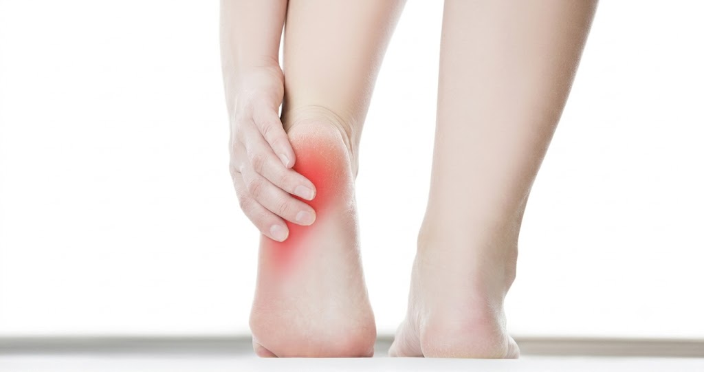
The classic location is beneath the heel, usually towards its inner or medial side where the central plantar fascia arises from the calcaneus. Some people feel pain extending a short distance into the arch.
Typical features include:
- pain during the first steps after sleep or rest;
- tenderness at the inner plantar heel;
- pain after prolonged standing or walking;
- symptoms following a recent increase in weight-bearing activity;
- pain when the toes are bent upwards and the fascia is tensioned; and
- a pattern that may ease after warming up but worsen again with accumulated load.
The pain may be described as sharp, pulling, aching or bruised. The description alone cannot confirm the diagnosis. Location, timing, provoking activities and examination findings must fit together.
Who develops plantar fasciitis?
Plantar fasciitis affects runners, walkers, people who stand at work, people returning to exercise and people with relatively low activity levels. Estimates commonly suggest that plantar heel pain affects approximately 4–7% of the community, while the often-quoted lifetime estimate for plantar fasciitis is around 10%. These figures vary because studies use different definitions and populations (Morrissey et al., 2021; Tan et al., 2024).
A multidisciplinary Singapore consensus guideline for plantar fasciitis was published in 2024. That paper noted that local prevalence was still being investigated, so it would be misleading to claim that a precise percentage of Singaporeans has the condition (Tan et al., 2024).
Evidence has associated plantar fasciitis or chronic plantar heel pain with:
- limited ankle dorsiflexion, particularly when the calf-Achilles complex restricts forward movement of the leg over the foot;
- prolonged occupational weight-bearing;
- a recent increase in weight-bearing or running load;
- higher body mass index in non-athletic populations; and
- reduced foot and ankle muscle capacity in some patients.
A classic matched case-control study found associations with reduced ankle dorsiflexion, higher body mass index and spending much of the workday on the feet (Riddle et al., 2003). Reviews have supported the association between higher body mass index and plantar fasciopathy, especially in non-athletic groups (van Leeuwen et al., 2016). Among physically active people, the overall risk-factor literature remains limited and does not justify treating every proposed biomechanical variable as a proven cause (Hamstra-Wright et al., 2021).
Body size should never be used to blame a patient. It changes mechanical demand, but it does not provide a complete explanation: many people in larger bodies have no heel pain, while many lean athletes develop plantar fasciitis.
Flat feet, high arches and pronation
Both flatter and higher-arched feet are frequently listed as risk factors. The relationship is not simple.
Pronation is a normal movement that helps the foot adapt and absorb load. A flatter-looking foot is not automatically abnormal, and a high arch is not automatically protective. What may matter is how far, how fast and for how long a foot moves; how the rest of the limb behaves; the loads involved; and whether the tissues tolerate those loads.
Systematic evidence has not consistently supported every traditional claim about static foot posture or ankle mechanics (van Leeuwen et al., 2016). A clinician should therefore assess foot posture as one part of a complete examination, not diagnose the cause of heel pain by looking at the arch alone.
Is plantar fasciitis especially relevant in Singapore?
Singapore does not have a unique form of plantar fasciitis. However, local routines can create relevant combinations of load:
- prolonged standing in healthcare, education, retail, hospitality and other occupations;
- frequent walking on pavement, tiled floors and hard indoor surfaces;
- sudden increases in walking during travel;
- transitions between office shoes, sandals, slippers and sports shoes; and
- bursts of running, court sport, gym training or hiking around otherwise sedentary working weeks.
These factors do not prove that hard floors, tropical footwear or city living independently cause plantar fasciitis. They may modify the demands placed on an already sensitive structure.
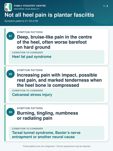
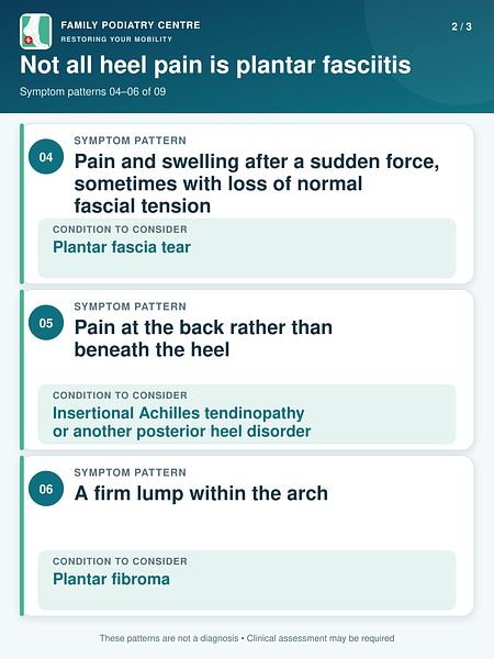
Not all heel pain is plantar fasciitis
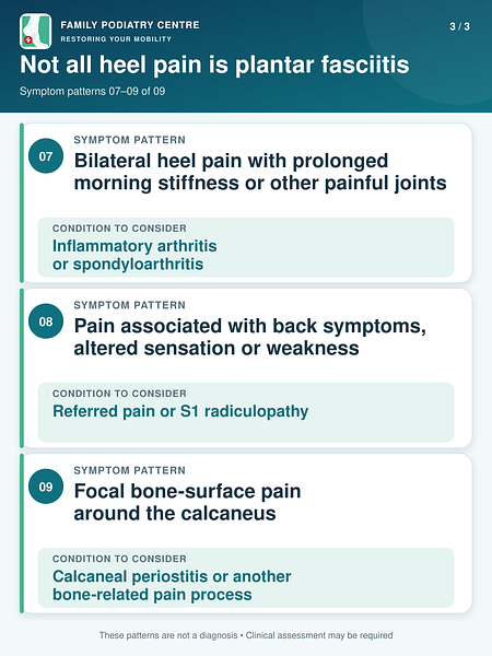
“Plantar heel pain” describes a location, not one guaranteed diagnosis. Several tissues share a small anatomical area around the heel. Treating all pain beneath the heel as plantar fasciitis can delay the correct care.
The clinical guideline specifically advises considering alternative diagnoses when the pattern is inconsistent with plantar fasciitis or symptoms fail to respond as expected (Koc
et al., 2023). For deeper information on two commonly confused conditions, see our guides to
heel fat pad pain and
calcaneal periostitis.
When heel pain needs prompt medical assessment
Arrange timely professional assessment if heel pain:
- followed a significant injury or sudden tearing sensation;
- prevents normal weight-bearing;
- is accompanied by marked swelling, redness, warmth, fever or an open wound;
- is associated with numbness, progressive weakness or altered foot control;
- is severe at rest or repeatedly wakes you at night;
- occurs with unexplained systemic symptoms;
- affects a person with diabetes, impaired circulation, neuropathy or immune suppression; or
- is worsening rather than stabilising despite sensible load reduction.
These findings do not automatically indicate a serious disorder, but they justify a different level of investigation.
Book An AppointmentHow is plantar fasciitis diagnosed?
In a typical case, plantar fasciitis is a clinical diagnosis. A detailed history and physical examination are usually more useful than ordering imaging immediately (Koc et al., 2023; Tan et al., 2024).
The history
The clinician should ask:
- Where exactly is the pain?
- Is it worst on the first steps after rest?
- Does it improve with brief movement and return after prolonged load?
- Was there a recent change in work, walking, running, sport, travel or footwear?
- Are there neurological symptoms, night pain, swelling or systemic symptoms?
- What treatments have already been attempted, and how did the symptoms respond?
The physical examination
A useful examination may include:
- palpating the plantar fascia attachment and other parts of the heel;
- assessing ankle dorsiflexion and calf flexibility;
- performing the windlass test by tensioning the plantar fascia through the big toe;
- screening the heel bone, fat pad, Achilles tendon and nerves;
- evaluating foot posture and joint movement;
- testing calf, foot and ankle strength;
- observing standing, walking and, where relevant, running; and
- assessing footwear and occupational or sporting demands.
Video gait analysis and plantar-pressure measurement may help identify how an individual loads the heel, arch and forefoot. They can guide mechanical treatment, footwear advice and orthotic design. They do not, by themselves, prove that pain comes from the plantar fascia. Their findings must be interpreted with the history and clinical examination.
Should pain be measured?
Yes. A simple daily score for first-step pain and a measure of function can make progress more visible. Clinical guidelines recommend validated tools such as the Foot Function Index, Foot and Ankle Ability Measure or Foot Health Status Questionnaire (Koc et al., 2023). In routine care, the same repeatable measure should be used over time rather than relying only on memory.
Do I need an X-ray, ultrasound or MRI?
Most patients with a classic presentation do not need imaging at the first visit.
Imaging becomes more useful when:
- the diagnosis is uncertain;
- symptoms are atypical or severe;
- a tear, stress injury, nerve disorder, inflammatory disease or other diagnosis is suspected; or
- an appropriate period of conservative care has not produced the expected progress.
Diagnostic ultrasound
Ultrasound can show plantar fascia thickness, texture, tears and changes around the heel attachment. A thickness greater than about 4 mm and a darker, hypoechoic appearance may support the diagnosis, but scan findings must be interpreted clinically because structural changes can occur in people without pain (McMillan et al., 2009; Koc et al., 2023; Drake et al., 2022).
Singapore’s 2024 consensus guideline supports bedside ultrasound as a useful tool to confirm the diagnosis when available, while also stating that it is not essential for every straightforward case (Tan et al., 2024).
Diagnostic ultrasound should not be confused with therapeutic ultrasound, a passive treatment in which sound energy is applied to the tissue. The 2023 guideline advises against using therapeutic ultrasound merely to enhance a stretching programme, and a 2025 meta-analysis found no convincing reduction in pain from ultrasound alone or when added to conventional exercise (Koc et al., 2023; Li et al., 2025).
X-rays:
X-rays show bone, not the health of the plantar fascia. They may be appropriate when a bone problem is suspected or in persistent, unexplained pain. Finding a heel spur does not prove that the spur is the source of symptoms.
MRI:
MRI can assess fascia, bone marrow, nerves and other soft tissues in greater detail. It is usually reserved for atypical, complicated or unresolved cases rather than routine plantar fasciitis.
Does a heel spur cause plantar fasciitis?
A plantar heel spur is a bony projection from the calcaneus. Spurs are associated with plantar heel pain at a population level, but association is not the same as causation. Many people have spurs without pain, and many painful heels do not require the spur to be removed (Kirkpatrick, Yassaie and Mirjalili, 2017).
In a long-term cohort of people with severe plantar fasciitis, the presence of a heel spur at baseline did not predict whether symptoms persisted (Hansen et al., 2018). The pain-generating system is usually more complex than a piece of bone “stabbing” the foot.
Treatment should therefore focus on the clinical source of pain and the factors maintaining it—not automatically on eliminating the spur.
Evidence-based treatment: what should happen first?
No single treatment works for every patient. The most defensible approach is a core programme followed by stepped, individualised care.
Comparative research has not established one intervention as universally superior, reinforcing the need to match care to the diagnosis, presentation and response over time (Babatunde et al., 2019).
The 2021 best-practice guide combined systematic review evidence, expert clinical reasoning and patient experience. It recommended plantar fascia stretching, taping and individualised education as the core approach, with footwear advice included. If progress is inadequate, shockwave therapy and then orthoses may be considered within stepped care (Morrissey et al., 2021). A 2026 topical review retains the same broad core approach (Cotchett and Morrissey, 2026).
The 2023 clinical practice guideline gives strong recommendations to stretching, taping and targeted manual therapy, supports foot and ankle resistance exercise, recommends night splints for consistent first-step pain and advises that orthoses should be combined with other treatments rather than used in isolation (Koc et al., 2023).
Singapore’s guideline similarly places education, footwear advice, activity modification and stretching within standard care and provides a pathway for escalation in persistent cases (Tan et al., 2024).
1. Education and realistic expectations
Understanding the condition is treatment, not an optional extra. Patients need to know:
- the fascia is painful but is not necessarily permanently damaged;
- recovery usually takes longer than a few days;
- complete rest can reduce capacity and is rarely the long-term answer;
- continuing exactly the same aggravating load can prevent improvement;
- a good plan adjusts load while progressively restoring capacity; and
- symptom change should guide progression.
This avoids the two unhelpful extremes of “ignore the pain and push through” and “never use the foot until all pain disappears.”
2. Modify load without becoming inactive
Load modification means temporarily reducing the activities that produce the largest or most prolonged symptom increase. Depending on the person, that may mean:
- shorter walks with planned breaks;
- fewer consecutive standing hours;
- temporarily reducing running distance, speed, hills or jumping;
- substituting a better-tolerated form of exercise;
- avoiding sudden increases in step count; or
- rotating tasks at work where possible.
A useful monitoring principle is the 24-hour response. Mild discomfort during activity may be acceptable for some patients if it settles promptly and the next morning’s first-step pain is not meaningfully worse. A substantial or sustained next-day increase suggests that the dose exceeded current capacity. This is a monitoring principle, not a universal pain threshold; acute tears, bone stress injuries and other diagnoses require different rules.
3. Plantar fascia-specific stretching
Plantar fascia-specific stretching has direct trial support and is strongly recommended in current guidance (DiGiovanni et al., 2003; DiGiovanni et al., 2006; Koc et al., 2023).
A commonly used method is:
- Sit and cross the affected foot over the other leg.
- Hold the toes, especially the big toe, and gently pull them towards the shin.
- Use the other hand to feel for tension along the plantar fascia in the arch.
- Hold a gentle, tolerable stretch without forcing into sharp pain.
- Repeat before the first steps in the morning and after prolonged sitting.
The original research protocol used repeated short holds. Exact dosage should be adapted to irritability, age, mobility and the diagnosis. More force is not automatically better.
4. Calf stretching
The gastrocnemius and soleus influence ankle dorsiflexion and how the body progresses over the foot. Current guidelines recommend stretching both the plantar fascia and the calf complex (Koc et al., 2023; Tan et al., 2024).
Two positions are commonly used:
- Knee straight: biases the gastrocnemius.
- Knee slightly bent: biases the soleus.
The heel should remain in contact with the ground and the stretch should be felt in the calf, not as sharp pain under the heel. Stretching should not be used to force a painful tissue beyond tolerance.
5. Taping
Rigid Low-Dye taping or an elastic taping technique can reduce pain and improve function over the short term when used with other care. Taping may also act as a clinical test: if symptoms improve when the foot is temporarily supported, longer-term mechanical support may be worth considering (Koc et al., 2023).
An updated 2026 systematic review found that Low-Dye taping improved pain and quality of life compared with placebo, although it was not superior to every other treatment (Zhang et al., 2026). Taping is best understood as a temporary load-modifying tool, not a cure. Skin sensitivity, circulation and tape tolerance must be considered.
6. Progressive strengthening
Stretching can reduce symptoms, but the longer-term aim is also to increase the capacity of the foot and ankle system.
The 2023 guideline recommends resistance training for the foot and ankle muscles (Koc et al., 2023). A randomised trial found faster improvement at three months with a progressive high-load heel-raise programme than with plantar fascia stretching alone, although outcomes were similar later in follow-up (Rathleff et al., 2015).
Strengthening may include appropriately progressed:
- calf raises;
- bent-knee calf work;
- toe-flexor and intrinsic-foot exercise;
- inversion and eversion resistance; and
- balance and lower-limb control exercises.
High-load exercise is not the correct starting point for every painful heel. The dose should match symptom irritability and current strength. A patient who cannot tolerate ordinary walking without a prolonged flare needs a different entry point from an athlete with mild, stable morning pain.
7. Footwear
Shoes can change heel pressure, comfort, stability and the way forces move through the foot, but no brand or model is a universal plantar fasciitis treatment.
Useful characteristics may include:
- adequate fit in length, width and depth;
- a stable platform appropriate to the individual;
- sufficient heel cushioning without excessive instability;
- a sole that does not demand painful movement at the wrong time; and
- enough internal volume for an orthosis if one is prescribed.
A 2025 laboratory study in women with plantar heel pain found that shoe and insert design could alter heel pressure, loading rate and comfort. It was an immediate-effects study, however, not proof that one commercial shoe cures the condition (Franettovich Smith et al., 2025).
Barefoot walking is not inherently unhealthy. During an irritable episode, however, repeated barefoot walking on hard flooring may be less comfortable because there is less external cushioning or load modification. Temporary indoor footwear can be helpful for some patients. The response should be assessed individually.
8. Foot orthoses
Foot orthoses may cushion the heel, modify arch deformation, redistribute plantar pressure or alter the timing and magnitude of load. Their value depends on the patient, the device and the role they play within a broader programme.
The evidence requires a balanced interpretation:
- orthoses may provide modest pain improvement, particularly over the medium term;
- prefabricated and custom devices can both help;
- evidence does not show that custom orthoses are universally superior to prefabricated devices;
- orthoses should not be presented as a stand-alone cure; and
- customisation may be clinically valuable when foot shape, force pattern, footwear, activity, deformity, pressure sensitivity or failure of a suitable prefabricated device justifies it.
An earlier randomised trial found small short-term benefits and broadly similar outcomes between customised and prefabricated devices (Landorf, Keenan and Herbert, 2006). Systematic reviews have found medium-term pain benefit but uncertainty about the size and consistency of the effect (Whittaker et al., 2018). The 2023 guideline explicitly advises against using either prefabricated or custom orthoses as an isolated short-term treatment, while allowing them as part of combined care (Koc et al., 2023). A newer review similarly found pain improvement without clearly meaningful improvement in every functional outcome (Wang et al., 2025).
The correct question is not “Are custom orthotics always best?” It is “Does this patient need an orthosis, what mechanical goal should it serve, and how will we confirm that it is helping?”
9. Night splints
A night splint holds the ankle and toes in a position intended to reduce overnight shortening of the calf–plantar fascia system. The 2023 guideline recommends a one- to three-month programme for patients who consistently experience first-step morning pain (Koc et al., 2023).
Night splints are not equally comfortable or necessary for everyone. Fit, sleep disruption, nerve symptoms and adherence matter. They are more logical when post-rest pain is a prominent and persistent feature.
10. Manual therapy
Targeted manual therapy may help when examination identifies relevant joint restriction, calf or soft-tissue limitation. Guidelines support manual therapy directed at lower-limb joints and soft tissues to address identified restrictions and improve pain and function (Fraser et al., 2018; Koc et al., 2023).
This does not justify forceful, painful massage directly into every sore heel. Aggressive pressure that produces a prolonged flare is not a sign of successful treatment. Gentle self-massage may feel helpful to some people, while clinician-applied techniques should have a defined objective and be monitored by response.
11. Ice, heat and pain medication
Ice may provide temporary symptom relief after an aggravating period, but it does not correct the mechanical or capacity-related contributors. Heat may feel soothing for stiffness in some patients. Neither should replace active management.
Pain-relieving or anti-inflammatory medication may be appropriate for selected patients, but chronic plantar fasciopathy is not simply an inflammatory disorder. Medication choice must consider age, allergies, pregnancy, kidney and gastrointestinal health, cardiovascular risk and interactions with other medicines. A pharmacist or doctor should advise when there is uncertainty.
Book An AppointmentWhat if the pain is not improving?
Before adding increasingly invasive treatment, reassess the basics:
- Is the diagnosis correct?
- Has the activity dose actually changed?
- Is the home programme being performed as intended?
- Is the exercise dose too high, too low or poorly progressed?
- Are work and footwear repeatedly recreating the aggravating load?
- Is there a stress injury, fat pad problem, nerve disorder, fascia tear or inflammatory condition?
- Is progress being judged with repeatable outcome measures?
Persistent pain is not automatically treatment failure. It may reveal a missed diagnosis, an unaddressed load or a programme that does not match the patient.
Extracorporeal shockwave therapy
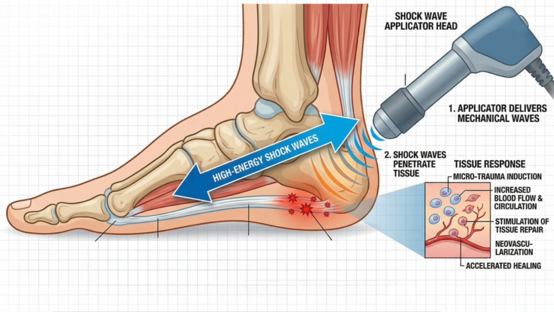
Extracorporeal shockwave therapy (ESWT) applies mechanical acoustic pulses to the painful region. It is non-surgical and does not involve injecting a drug. The precise biological effects remain under investigation, but may include altered pain signalling and stimulation of tissue-remodelling pathways.
Evidence supports ESWT as an option for persistent plantar fasciopathy, although treatment parameters vary across studies. A 2024 systematic review found it effective and generally tolerable but noted that energy, pulse number and protocol differences can influence results (Lippi et al., 2024). Singapore’s consensus guideline places ESWT among the options for recalcitrant plantar fasciitis before corticosteroid injection or surgery (Tan et al., 2024).
Shockwave should still sit within a complete plan. It does not remove the need to manage load, restore strength and address relevant footwear or mechanical factors.
Corticosteroid injection
Corticosteroid injection may reduce pain over a short period for some patients, but the evidence is less impressive when compared with placebo and when lower-quality studies are excluded. A systematic review found no clear superiority over placebo for pain or function, despite advantages over some active comparators in certain time windows (Whittaker et al., 2019).
Potential risks include post-injection flare, infection, nerve injury, fat-pad injury and plantar fascia rupture. Absolute risk is difficult to quantify from trials because uncommon adverse events require large samples and long follow-up. If corticosteroid is considered, diagnosis, technique, injection location, ultrasound guidance, prior injections and the post-injection loading plan all matter.
Singapore guidance places corticosteroid later in the pathway for persistent cases rather than as a routine first response (Tan et al., 2024).
Platelet-rich plasma and other injections
Platelet-rich plasma (PRP) uses a concentrated component of the patient’s own blood. Some reviews report better medium- or longer-term outcomes than corticosteroid, but study quality, preparation methods, injection protocols and comparison treatments vary substantially (Hohmann, Tetsworth and Glatt, 2021).
A large 2026 network meta-analysis compared multiple minimally invasive approaches, including ESWT and several injection types. It illustrates that some interventions can outperform placebo for selected outcomes, but comparisons are constrained by inconsistent trials and protocols (Tien et al., 2026). PRP, prolotherapy, botulinum toxin and other injections should therefore not be marketed as universally proven cures.
Surgery
Surgery is uncommon and reserved for carefully selected, persistent cases after an adequate course of appropriate conservative care. Options may include partial plantar fascia release or gastrocnemius procedures when a clinically important calf contracture is present.
Singapore consensus guidance allows surgery to be considered when symptoms have persisted for more than six months and conservative treatment has failed (Tan et al., 2024). In practice, the decision also depends on diagnostic certainty, severity, functional loss, previous treatment quality and the risks of altering arch mechanics, nerve function or scar sensitivity.
How long does plantar fasciitis take to heal?
There is no reliable promise that applies to everyone. Many patients improve over several months with conservative care, but a minority have symptoms for much longer.
An often-cited long-term study found that symptoms persisted in a substantial proportion of participants. That result requires context: the cohort had severe, difficult-to-treat plantar fasciitis and therefore should not be used to predict the outcome of every new case. Even among those who became symptom-free, mean symptom duration was approximately two years (Hansen et al., 2018).
The most useful message is neither “it will disappear in a few days” nor “you will have it forever.” Recovery is variable. A correct diagnosis, early load adjustment, consistent core care and progressive restoration of capacity give the tissue a better environment in which to improve.
Signs of genuine progress
Progress is more than a lower pain score at one moment. Useful signs include:
- less first-step pain;
- fewer painful first steps;
- reduced tenderness at the plantar fascia attachment;
- longer comfortable standing or walking time;
- improved calf and foot strength;
- less next-day reaction to activity; and
- gradual return to work, exercise or sport without repeated setbacks.
Imaging does not need to return to “normal” before a patient can recover. Structural abnormalities may remain after symptoms resolve (Hansen et al., 2018).
Can I keep walking or running?
Many people can remain active with a modified dose. Whether walking or running is appropriate depends on the diagnosis, symptom irritability, gait, next-day response and the demands of the activity.
Running may be continued or reintroduced when:
- ordinary walking is well tolerated;
- morning pain is stable or improving;
- the patient can perform appropriate calf-loading tasks without a sustained flare;
- running volume and intensity can be controlled; and
- symptoms return to baseline by the agreed monitoring period.
Distance, speed, hills and consecutive running days should not all increase at once. Bone stress injury, acute fascia tear and neurological weakness require a different approach and should not be managed with a generic “run through it” rule.
A practical home-care framework
The following is educational rather than an individual prescription:
During a painful flare
- Reduce the largest aggravating loads for several days.
- Avoid long, unbroken periods of standing when possible.
- Use comfortable, stable footwear; temporary indoor cushioning may help.
- Perform gentle plantar fascia and calf stretching if it does not create a prolonged flare.
- Use ice for brief symptom relief if comfortable.
- Record first-step pain and the previous day’s load.
As symptoms stabilise
- Restore normal walking progressively.
- Begin or progress foot and ankle strengthening at a tolerable dose.
- Use taping or an orthosis if clinically indicated.
- Increase only one major activity variable at a time.
- Reassess if progress stalls or the pain pattern changes.
Do not use this framework to self-manage severe trauma, neurological symptoms, suspected stress injury, infection or a diabetic foot problem.
Common mistakes that delay recovery
Assuming every heel pain is plantar fasciitis
The wrong diagnosis leads to the wrong loading plan. Central heel pain, burning pain, bone pain and fascia pain should not be treated as interchangeable.
Chasing the heel spur
A spur can be present without causing pain. Treating the X-ray rather than the patient can lead to unnecessary fear and intervention.
Complete rest for too long
Brief relative rest can settle a flare. Prolonged avoidance can reduce calf, foot and tissue capacity, making return to activity harder.
Repeating an exercise that causes a sustained flare
Exercises require the correct dose. Pain that remains substantially worse the next day is feedback that the current dose or technique may be unsuitable.
Believing one shoe or insert will cure everyone
Foot shape, body size, work, sport, gait and comfort vary. Footwear and orthoses should serve a defined purpose within combined care.
Relying only on passive treatment
Shockwave, manual therapy, taping and pain relief may help, but long-term recovery usually also requires the patient to manage load and restore capacity.
Preventing recurrence
Not every episode can be prevented, but risk may be reduced by:
- increasing walking and running load gradually;
- maintaining calf and foot strength;
- avoiding large, abrupt changes in training, work or daily steps;
- replacing worn footwear when its structure no longer performs as intended;
- using footwear suited to the activity and individual;
- responding early to rising first-step pain; and
- maintaining the loading strategies that helped after symptoms settle.
Prevention is not about making the foot permanently dependent on support. It is about keeping demand, capacity and recovery in a sustainable relationship.
When should I see a podiatrist for plantar fasciitis?
Consider a professional assessment when:
- the diagnosis is uncertain;
- pain is severe, worsening or changing in character;
- heel pain is limiting work, walking, exercise or sleep;
- self-care has not produced clear progress;
- symptoms recur repeatedly;
- you have diabetes, neuropathy, inflammatory disease or circulatory problems;
- you need guidance on returning to running or sport; or
- footwear or orthoses need to be selected for a specific mechanical objective.
A podiatry assessment should establish what tissue is painful, why it is being overloaded, what the patient needs to return to and how progress will be measured. It should not begin with the assumption that every patient needs the same shoe, injection or orthotic.
At Family Podiatry Centre, plantar heel pain assessment combine clinical examination with video gait analysis and plantar-pressure measurement and other tools as are relevant. Findings are used to guide an individual plan that may include education, load modification, exercise, footwear changes, taping, orthoses or shockwave therapy.
Treatment selection depends on the diagnosis and the patient, not on a fixed package.
The central message
Plantar fasciitis is not simply inflammation, a heel spur, a flat foot or the consequence of one “bad” shoe. It is usually a multifactorial plantar fascia pain disorder in which tissue loading and tissue capacity have fallen out of balance.
Good treatment begins with the correct diagnosis. It then combines education, sensible load modification, stretching, progressive strengthening and appropriate mechanical support. Taping, footwear changes, orthoses, night splints, manual therapy and shockwave can each have a role, but their value depends on the patient and where they fit within the complete plan.
That is a more honest message than promising an instant cure. It is also a more useful one: most painful heels can improve, but the route to improvement should be measured, progressive and specific to the person.
Book An Appointment
Disclaimer: The word "treatment" in this article refers to the care and management of a patient’s health to prevent, cure, or improve a condition. Treatment results vary and do not necessarily indicate a cure. This article is for informational and educational purposes only and does not constitute medical advice.
About the AuthorMark B. Reyneker, BTech (Podiatry), MSc (Palaeontology) is a podiatrist and Founder & Clinical Director of Family Podiatry Centre, with more than 25 years of clinical experience across South Africa, Malaysia and Singapore. His clinical interests include foot and lower-limb pain, gait and biomechanics, sports-related foot conditions, orthotic therapy and footwear.
Alongside his clinical practice, Mark conducts research into human gait and foot biomechanics. His MSc research at the University of the Witwatersrand investigated human propulsion and the structural properties of the metatarsals. He is also the inventor of
A Foot Orthotic, an orthotic technology developed through an international patent family.
References
Babatunde, O.O., Legha, A., Littlewood, C., Chesterton, L.S., Thomas, M.J., Menz, H.B., van der Windt, D. and Roddy, E. (2019) ‘Comparative effectiveness of treatment options for plantar heel pain: a systematic review with network meta-analysis’,
British Journal of Sports Medicine, 53(3), pp. 182–194. Available at:
https://doi.org/10.1136/bjsports-2017-098998.
DiGiovanni, B.F., Nawoczenski, D.A., Lintal, M.E., Moore, E.A., Murray, J.C., Wilding, G.E. and Baumhauer, J.F. (2003) ‘Tissue-specific plantar fascia-stretching exercise enhances outcomes in patients with chronic heel pain: a prospective, randomized study’,
Journal of Bone and Joint Surgery. American Volume, 85(7), pp. 1270–1277. Available at:
https://doi.org/10.2106/00004623-200307000-00013.
DiGiovanni, B.F., Nawoczenski, D.A., Malay, D.P., Graci, P.A., Williams, T.T., Wilding, G.E. and Baumhauer, J.F. (2006) ‘Plantar fascia-specific stretching exercise improves outcomes in patients with chronic plantar fasciitis: a prospective clinical trial with two-year follow-up’,
Journal of Bone and Joint Surgery. American Volume, 88(8), pp. 1775–1781. Available at:
https://doi.org/10.2106/JBJS.E.01281.
Drake, C., Whittaker, G.A., Kaminski, M.R., Chen, J., Keenan, A.-M., Rathleff, M.S., Robinson, P. and Landorf, K.B. (2022) ‘Medical imaging for plantar heel pain: a systematic review and meta-analysis’,
Journal of Foot and Ankle Research, 15, 4. Available at:
https://doi.org/10.1186/s13047-021-00507-2.
Erdemir, A., Hamel, A.J., Fauth, A.R., Piazza, S.J. and Sharkey, N.A. (2004) ‘Dynamic loading of the plantar aponeurosis in walking’,
Journal of Biomechanics, 37(11), pp. 1643–1649. Available at:
https://doi.org/10.1016/j.jbiomech.2004.02.029.
Franettovich Smith, M.M., van den Hoorn, W., van den Hoek, A., Kerr, G.K. and Hurn, S.E. (2025) ‘Immediate effects of footwear design on in-shoe plantar pressures, impact forces and comfort in women with plantar heel pain’,
Journal of Foot and Ankle Research, 18(2), e70055. Available at:
https://doi.org/10.1002/jfa2.70055.
Fraser, J.J., Corbett, R., Donner, C. and Hertel, J. (2018) ‘Does manual therapy improve pain and function in patients with plantar fasciitis? A systematic review’,
Journal of Manual & Manipulative Therapy, 26(2), pp. 55–65. Available at:
https://doi.org/10.1080/10669817.2017.1322736.
Hamstra-Wright, K.L., Huxel Bliven, K.C., Bay, R.C. and Aydemir, B. (2021) ‘Risk factors for plantar fasciitis in physically active individuals: a systematic review and meta-analysis’,
Sports Health, 13(3), pp. 296–303. Available at:
https://doi.org/10.1177/1941738120970976.
Hansen, L., Krogh, T.P., Ellingsen, T., Bolvig, L. and Fredberg, U. (2018) ‘Long-term prognosis of plantar fasciitis: a 5- to 15-year follow-up study of 174 patients with ultrasound examination’,
Orthopaedic Journal of Sports Medicine, 6(3), 2325967118757983. Available at:
https://doi.org/10.1177/2325967118757983.
Hohmann, E., Tetsworth, K. and Glatt, V. (2021) ‘Platelet-rich plasma versus corticosteroids for the treatment of plantar fasciitis: a systematic review and meta-analysis’,
American Journal of Sports Medicine, 49(5), pp. 1381–1393. Available at:
https://doi.org/10.1177/0363546520937293.
Kirkpatrick, J., Yassaie, O. and Mirjalili, S.A. (2017) ‘The plantar calcaneal spur: a review of anatomy, histology, etiology and key associations’,
Journal of Anatomy, 230(6), pp. 743–751. Available at:
https://doi.org/10.1111/joa.12607.
Koc, T.A. Jr, Bise, C.G., Neville, C., Carreira, D., Martin, R.L. and McDonough, C.M. (2023) ‘Heel pain—plantar fasciitis: revision 2023’,
Journal of Orthopaedic & Sports Physical Therapy, 53(12), pp. CPG1–CPG39. Available at:
https://doi.org/10.2519/jospt.2023.0303.
Landorf, K.B., Keenan, A.-M. and Herbert, R.D. (2006) ‘Effectiveness of foot orthoses to treat plantar fasciitis: a randomized trial’,
Archives of Internal Medicine, 166(12), pp. 1305–1310. Available at:
https://doi.org/10.1001/archinte.166.12.1305.
Lemont, H., Ammirati, K.M. and Usen, N. (2003) ‘Plantar fasciitis: a degenerative process (fasciosis) without inflammation’,
Journal of the American Podiatric Medical Association, 93(3), pp. 234–237. Available at:
https://doi.org/10.7547/87507315-93-3-234.
Li, X., Yang, J., Li, S., Yang, L. and Meng, D. (2025) ‘The efficacy of ultrasound for plantar fasciitis: a systematic review and meta-analysis’,
Annals of Medicine, 57(1), 2543056. Available at:
https://doi.org/10.1080/07853890.2025.2543056.
Lippi, L., Folli, A., Moalli, S., Turco, A., Ammendolia, A., de Sire, A. and Invernizzi, M. (2024) ‘Efficacy and tolerability of extracorporeal shock wave therapy in patients with plantar fasciopathy: a systematic review with meta-analysis and meta-regression’,
European Journal of Physical and Rehabilitation Medicine, 60(5), pp. 832–846. Available at:
https://doi.org/10.23736/S1973-9087.24.08136-X.
McMillan, A.M., Landorf, K.B., Barrett, J.T., Menz, H.B. and Bird, A.R. (2009) ‘Diagnostic imaging for chronic plantar heel pain: a systematic review and meta-analysis’,
Journal of Foot and Ankle Research, 2, 32. Available at:
https://doi.org/10.1186/1757-1146-2-32.
Morrissey, D., Cotchett, M., Said J’Bari, A., Prior, T., Griffiths, I.B., Rathleff, M.S., Gulle, H., Vicenzino, B. and Barton, C.J. (2021) ‘Management of plantar heel pain: a best practice guide informed by a systematic review, expert clinical reasoning and patient values’,
British Journal of Sports Medicine, 55(19), pp. 1106–1118. Available at:
https://doi.org/10.1136/bjsports-2019-101970.
Rathleff, M.S., Mølgaard, C.M., Fredberg, U., Kaalund, S., Andersen, K.B., Jensen, T.T., Aaskov, S. and Olesen, J.L. (2015) ‘High-load strength training improves outcome in patients with plantar fasciitis: a randomized controlled trial with 12-month follow-up’,
Scandinavian Journal of Medicine & Science in Sports, 25(3), pp. e292–e300. Available at:
https://doi.org/10.1111/sms.12313.
Riel, H., Cotchett, M., Delahunt, E., Rathleff, M.S., Vicenzino, B., Weir, A. and Landorf, K.B. (2017) ‘Is “plantar heel pain” a more appropriate term than “plantar fasciitis”? Time to move on’,
British Journal of Sports Medicine, 51(22), pp. 1576–1577. Available at:
https://doi.org/10.1136/bjsports-2017-097519.
Riddle, D.L., Pulisic, M., Pidcoe, P. and Johnson, R.E. (2003) ‘Risk factors for plantar fasciitis: a matched case-control study’,
Journal of Bone and Joint Surgery. American Volume, 85(5), pp. 872–877. Available at:
https://doi.org/10.2106/00004623-200305000-00015.
Tan, V.A.K., Tan, C.C., Yeo, N.E.M., Zhang, M., Mehta, K.V., Ho, R.H.T. and Tan, B. (2024) ‘Consensus statements and guideline for the diagnosis and management of plantar fasciitis in Singapore’,
Annals of the Academy of Medicine, Singapore, 53(2), pp. 101–112. Available at:
https://doi.org/10.47102/annals-acadmedsg.2023211.
Tien, C.H., Chiu, M.C., Shen, Y.L., Ko, Y.C. and Lee, J.J. (2026) ‘Comparative effectiveness of minimally invasive therapies for plantar fasciitis: a systematic review and network meta-analysis’,
Scientific Reports, 16, 9074. Available at:
https://doi.org/10.1038/s41598-026-40038-z.
van Leeuwen, K.D.B., Rogers, J., Winzenberg, T. and van Middelkoop, M. (2016) ‘Higher body mass index is associated with plantar fasciopathy/“plantar fasciitis”: systematic review and meta-analysis of various clinical and imaging risk factors’,
British Journal of Sports Medicine, 50(16), pp. 972–981. Available at:
https://doi.org/10.1136/bjsports-2015-094695.
Wang, D., Lin, Z., Tan, G., Han, X. and Huang, Y. (2025) ‘Efficacy and safety of foot orthoses for improving pain and function in patients with plantar fasciitis: a systematic review and meta-analysis of randomized controlled trials’,
Disability and Rehabilitation, pp. 1–15. Available at:
https://doi.org/10.1080/09638288.2025.2563763.
Whittaker, G.A., Munteanu, S.E., Menz, H.B., Tan, J.M., Rabusin, C.L. and Landorf, K.B. (2018) ‘Foot orthoses for plantar heel pain: a systematic review and meta-analysis’,
British Journal of Sports Medicine, 52(5), pp. 322–328. Available at:
https://doi.org/10.1136/bjsports-2016-097355.
Whittaker, G.A., Munteanu, S.E., Menz, H.B., Bonanno, D.R., Gerrard, J.M. and Landorf, K.B. (2019) ‘Corticosteroid injection for plantar heel pain: a systematic review and meta-analysis’,
BMC Musculoskeletal Disorders, 20, 378. Available at:
https://doi.org/10.1186/s12891-019-2749-z.
Zhang, L., Chen, L., Zhang, X., Chen, J. and Luo, Y. (2026) ‘Effectiveness of Low-Dye taping in the treatment of patients with plantar fasciitis: a systematic review and meta-analysis’,
Medicine, 105(8), e46770. Available at:
https://doi.org/10.1097/MD.0000000000046770.