Published: 8 October 2026 | Last reviewed: 19 October 2026
Sesamoiditis is pain arising from repetitive stress and inflammation around the small sesamoid bones beneath the big-toe joint. Most people have two sesamoids under each first metatarsal head: a medial or tibial sesamoid and a lateral or fibular sesamoid. These bones sit inside the tendon system beneath the first metatarsophalangeal joint and help the big toe function efficiently during walking, running and push-off. Because they repeatedly accept high forces, they can become painful from overload. Pain beneath the big-toe joint should not automatically be labelled sesamoiditis, however, because stress fractures, acute fractures, bipartite sesamoids, osteoarthritis, osteonecrosis and other conditions can produce very similar symptoms.
Sesamoiditis commonly produces tenderness directly underneath the first metatarsal head and pain when weight moves forwards onto the big toe. The underlying mechanical cause and the condition of the sesamoid itself need to be established before treatment is planned.
Sesamoiditis is a particular type of ball-of-foot pain
Pain in the ball of the foot is often described broadly as metatarsalgia.
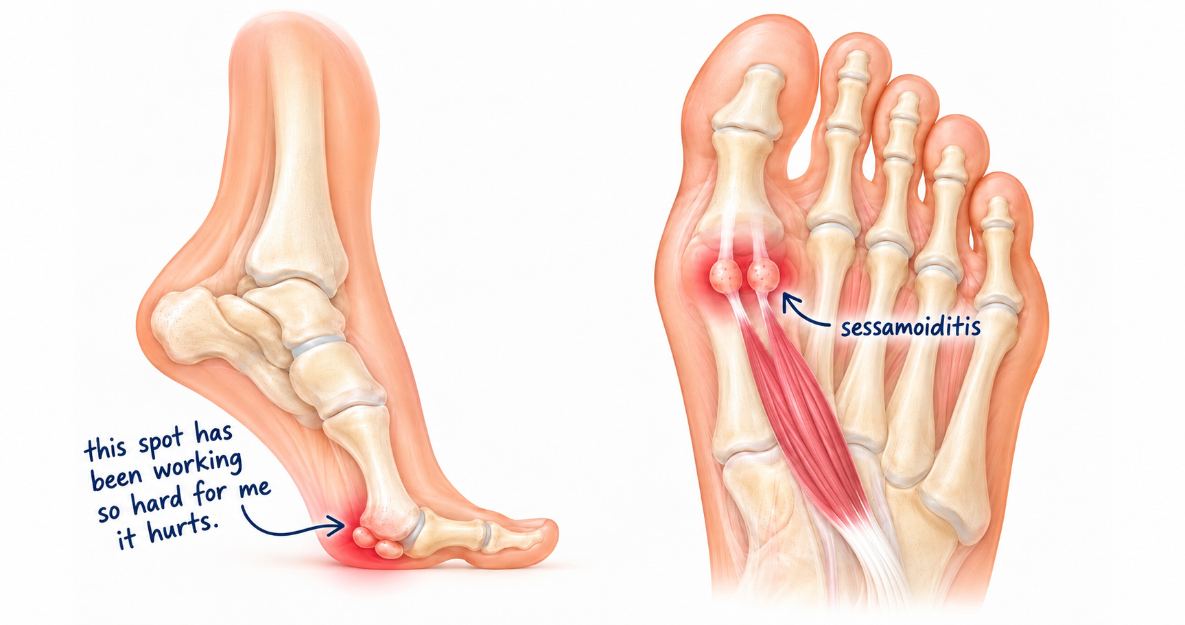
When the pain sits specifically underneath the big-toe joint, however, the anatomy is quite different from pain beneath the second, third or fourth metatarsal heads. Directly beneath the head of the first metatarsal sit two small bones. These are the hallucal sesamoids.
The word hallucal refers to the hallux, or big toe. The inner bone is usually called the medial or tibial sesamoid, while the outer one is called the lateral or fibular sesamoid. Despite their small size, they form an important part of the mechanical system responsible for moving the big toe and propelling the body forwards.
When this sesamoid complex becomes irritated by repeated loading, the resulting painful condition is commonly described as sesamoiditis.
Sesamoid bones are unusual because they develop inside tendons
Most bones form part of the structural skeleton and connect to other bones through joints. Sesamoids are different. They develop within tendons or closely associated soft-tissue structures at locations where mechanical forces are concentrated. The largest and best-known sesamoid bone in the human body is actually the patella, or kneecap.
The two sesamoids beneath the big toe perform a similar mechanical concept on a much smaller scale. They are embedded within the tendon apparatus of the flexor hallucis brevis, one of the muscles responsible for controlling the big toe. They are also intimately connected to the plantar capsule, plantar plate, collateral structures and surrounding tendons beneath the first metatarsophalangeal joint.
The long flexor tendon of the big toe — the flexor hallucis longus — passes between the two sesamoids. This means the sesamoids are not simply two spare bones sitting underneath the foot. They form part of an integrated pulley and stabilisation system. Anatomical studies describe their joint surfaces as being covered by cartilage where they articulate with the underside of the first metatarsal head. Ligaments connect the sesamoids to one another and to the surrounding joint complex.
The sesamoids act almost like a pulley underneath the big toe
One of their most important functions is mechanical. Muscles create movement by pulling through tendons. The effectiveness of that pull depends partly on the distance between the tendon and the centre of rotation of the joint. By holding the flexor tendon apparatus away from the first metatarsal head, the sesamoids help increase the mechanical advantage of the muscles controlling plantarflexion of the big toe.
In simple terms, they help the muscles underneath the big toe work more effectively. They also help protect the tendons passing through the region, distribute force underneath the first metatarsal head and provide a stable interface as body weight moves forwards over the foot. Research examining removal of a sesamoid has demonstrated measurable changes in the mechanical effectiveness of the flexor hallucis brevis, illustrating just how functionally important these small bones are. This becomes particularly relevant during propulsion.
As the heel lifts from the floor, body weight moves towards the front of the foot. The big-toe joint dorsiflexes while the first metatarsal head loads against the sesamoid complex underneath it. Walking exposes this system to repeated loading. Running increases that demand. Jumping, sprinting, dancing and activities involving repeated forefoot loading can increase it further.
The first metatarsal actually has grooves for the sesamoids
The relationship between the sesamoids and the metatarsal is remarkably specialised. On the plantar surface of the first metatarsal head are articular regions corresponding to the two sesamoids. Between them is an intersesamoidal ridge or crest. The sesamoids track on either side of this ridge as the big-toe joint moves. This arrangement helps maintain alignment between the first metatarsal, sesamoids and hallux during movement. It also explains why abnormalities of first-metatarsal position can influence the sesamoid complex.
In hallux valgus, for example, the metatarsal and sesamoids can progressively lose their normal relationship. Modern weight-bearing CT research has demonstrated that this is a three-dimensional problem involving translation and rotation rather than simply a big toe pointing sideways. The sesamoids therefore cannot be considered in isolation from the rest of the first ray.
The foot has 26 bones — except that it usually has more
One of the interesting inconsistencies in basic anatomy is the statement that the human foot contains 26 bones. The familiar calculation is: 7 tarsal bones + 5 metatarsals + 14 phalanges = 26 bones.
That calculation is correct as the standard skeletal count. But it usually leaves the two hallucal sesamoids out. If the two normal sesamoids beneath the big toe are included as separate ossified bones, the practical count becomes 28. And even 28 is not necessarily the absolute number of individual bones that can occur in every foot, because accessory ossicles and additional sesamoids can also occur.
The discrepancy exists because sesamoid and accessory bones have traditionally been treated differently from the main bones forming the axial framework of the foot. The standard 26 therefore describes the conventional structural skeleton rather than every separate piece of ossified tissue that may be present.
Leaving the hallucal sesamoids out of the bone count is anatomically convenient but slightly misleading
There is some logic behind the convention. Sesamoid bones form within tendons, and sesamoids elsewhere in the foot can be quite variable. Small sesamoids may occasionally occur beneath other metatarsophalangeal or interphalangeal joints, and numerous accessory ossicles can exist throughout the foot. Trying to include every anatomical variant would make the statement that the foot has a fixed number of bones impossible.
The two hallucal sesamoids, however, are somewhat different. They are remarkably consistent anatomical structures. Congenital absence has been described, but complete absence is extremely unusual. Anatomical reviews describe absent hallucal sesamoids as rare, while bilateral absence of both has been sufficiently unusual to warrant individual case reports in the medical literature.
In everyday clinical practice, therefore, seeing two sesamoids beneath the first metatarsal is overwhelmingly the expected anatomy. So when someone says there are 26 bones in a foot, it is useful to remember that the statement really means: 26 conventionally counted skeletal bones, plus the sesamoids and any accessory ossicles present.
Not every sesamoid is one single piece of bone
Another fascinating feature is that a sesamoid can develop in more than one ossification centre. Sometimes these centres remain separate. The result is a bipartite sesamoid.
Instead of one continuous bone, the sesamoid consists of two pieces. The medial or tibial sesamoid is much more commonly bipartite than the lateral sesamoid. Multipartite variants containing more than two components can also occur.
Published prevalence figures vary substantially between populations and imaging studies, but bipartite or multipartite hallucal sesamoids are well-recognised normal anatomical variants. This matters clinically because a bipartite sesamoid can sometimes resemble a fracture on an X-ray. The distinction is important.
A developmental separation commonly has smoother, more corticated margins, whereas an acute fracture may have a different appearance and an appropriate injury history. Occasionally the variant itself can also become painful. The mere presence of two pieces on an X-ray therefore does not automatically tell us what is causing the patient's pain.
Sesamoiditis usually develops through repeated loading rather than one dramatic injury
Sesamoiditis commonly develops gradually. A runner may notice increasing discomfort beneath the big-toe joint after longer distances. A person who spends prolonged periods standing may notice pain developing as the day progresses.
Another patient may experience symptoms after suddenly increasing exercise volume. Activities that repeatedly place the foot onto the forefoot can be particularly provocative. The tissue does not necessarily fail because one step was excessive. Instead, the amount of loading repeatedly reaching the sesamoid complex may exceed the ability of the area to recover between episodes of activity. This creates a load-versus-tissue-capacity problem.
Modern imaging literature describes sesamoiditis as a painful inflammatory response related to repetitive injury, with MRI potentially showing bone-marrow oedema and inflammation in adjacent soft tissues.
Pain is usually felt directly underneath the big-toe joint
The location is one of the most useful clues. Patients commonly point to the plantar surface underneath the first metatarsal head. There may be tenderness beneath one sesamoid more than the other. Walking barefoot on a hard floor may become particularly uncomfortable. Running can become painful.
Moving onto the toes, climbing stairs or performing movements that heavily load the forefoot may aggravate symptoms. Some patients describe a deep bruise-like sensation. Others describe sharper pain during push-off. The area may occasionally become swollen or inflamed. Because the sesamoids sit directly underneath the first metatarsal head, dorsiflexion of the big toe can also increase pressure or tension through the painful area.
Pain underneath a sesamoid does not automatically mean sesamoiditis
This distinction is critical. Several different disorders occur in almost exactly the same location.
A painful sesamoid may represent:
- repetitive inflammatory stress or sesamoiditis;
- a sesamoid stress reaction;
- a stress fracture;
- an acute traumatic fracture;
- a symptomatic bipartite sesamoid;
- osteoarthritis between the sesamoid and first metatarsal;
- osteonecrosis or avascular necrosis;
- injury to the surrounding plantar structures;
- pathology within the first metatarsophalangeal joint.
This is why sesamoiditis should not simply become a label for every pain underneath the big toe.
Recent literature on hallux sesamoid disorders emphasizes that sesamoid pain represents a group of different pathological conditions rather than one uniform problem. The correct diagnosis matters because the biological problem in an irritated but structurally intact sesamoid is not identical to the problem in a stress fracture or an osteonecrotic bone.
Sesamoid stress injury can initially feel very similar
Bone responds to repeated mechanical loading. Normally, this is positive. Bone continuously remodels in response to stress. Problems can develop when repetitive loading accumulates faster than the bone can remodel and recover. A bone stress reaction may develop before a visible fracture line exists.
If loading continues, this can progress towards a stress fracture. Early X-rays can therefore sometimes appear normal even when the patient has clinically significant bone stress. MRI is particularly useful in these situations because bone-marrow oedema may become visible before more obvious structural changes appear. This is one reason persistent focal pain underneath the first metatarsal should not simply be ignored as ordinary soreness.
An acute sesamoid fracture has a different history
Sesamoid fractures can also occur suddenly. A forceful landing, direct impact or abrupt loading event can injure the bone. The onset is usually more obvious than the gradual development typical of overuse-related sesamoid pain.
The distinction can nevertheless become difficult when a patient already has a bipartite sesamoid. Imaging then needs to be interpreted alongside the clinical history, location of tenderness and appearance of the bone margins. A picture on an X-ray should never be interpreted without knowing what happened to the patient.
Blood supply makes sesamoid disorders particularly interesting
These bones are small, but their vascular anatomy is important. Studies of the hallucal sesamoids describe blood vessels entering primarily from plantar and proximal directions, with relatively limited collateral circulation. This has been proposed as one factor contributing to problems such as delayed healing and osteonecrosis in certain sesamoid injuries.
Osteonecrosis, sometimes called avascular necrosis, occurs when bone viability is compromised because its blood supply becomes inadequate. It can produce chronic pain beneath the first metatarsal and may eventually cause changes in the structure of the sesamoid. The condition can resemble chronic sesamoiditis clinically, making imaging important when symptoms fail to follow the expected course.
The medial and lateral sesamoids do not always behave identically
The medial sesamoid lies beneath the inner side of the first metatarsal head and is frequently exposed to considerable load. It is also the sesamoid more commonly seen as a bipartite anatomical variant. The lateral sesamoid lies closer to the second metatarsal side of the first metatarsal head.
Although the two bones operate together, they have different soft-tissue attachments and slightly different mechanical environments. This is why an examination should identify which sesamoid is painful, rather than simply documenting generic pain beneath the big toe.
First-ray mechanics matter
A painful sesamoid is located at the end of a much larger mechanical chain. The first metatarsal, big toe, plantar fascia, sesamoids, flexor tendons and surrounding ligaments all interact during propulsion. Movement elsewhere in the foot can alter the pressure reaching this region.
The anatomy of the first ray, range of movement at the big-toe joint, alignment of the hallux, foot posture, activity demands and footwear can all influence loading. This does not mean that every patient with sesamoiditis has an obvious structural abnormality. It means the painful sesamoid should not be viewed as an isolated pebble underneath the foot. It is part of a complex moving joint.
Hallux valgus also changes the relationship between the sesamoids and first metatarsal
In a normally aligned first metatarsophalangeal joint, the sesamoids sit beneath the first metatarsal head within their corresponding grooves. Hallux valgus alters this relationship.
What often appears on a conventional X-ray as the sesamoids moving sideways is more accurately understood as a complex three-dimensional change involving movement and rotation of the first metatarsal relative to the sesamoid apparatus. Recent weight-bearing CT research has helped clarify this relationship.
This is important because pain beneath the first metatarsal in someone with hallux valgus may have a different mechanical background from the same pain in a runner with an otherwise straight big toe. Again, the location of pain alone does not explain its cause.
Examination aims to establish exactly which structure hurts
Assessment begins with precise localisation. Tenderness directly over the medial sesamoid is different from tenderness over the lateral sesamoid. Pain inside the first metatarsophalangeal joint may suggest another source. Movement of the big toe provides additional information.
The alignment of the first ray, mobility of the joint, surrounding soft tissues and loading pattern of the foot may also need consideration. The examination should then be interpreted alongside the patient's activity, recent changes in training, footwear and the way the symptoms began. A gradual overuse history creates a different diagnostic picture from sudden pain after landing heavily on the forefoot.
Imaging helps separate several conditions that feel almost identical
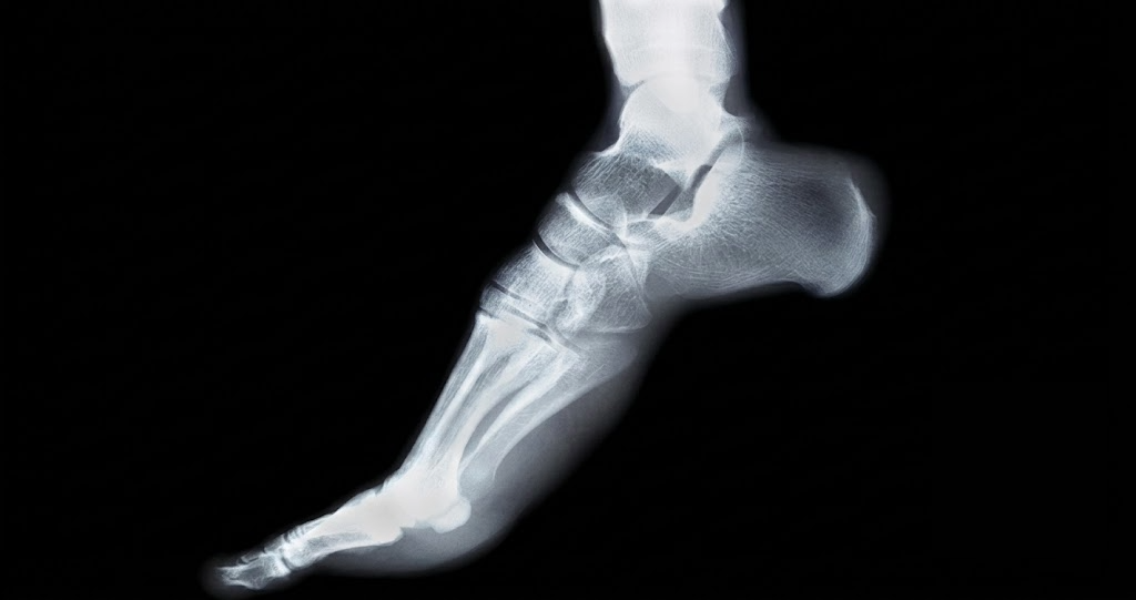
Plain radiographs remain important because they show the shape and structure of the sesamoids and first metatarsophalangeal joint. Specific sesamoid views can provide a tangential view of the two bones beneath the first metatarsal head.
These images may help demonstrate fractures, multipartite anatomy, degenerative change, alignment and some forms of chronic pathology. They also allow comparison of the sesamoids with the first-metatarsal articular surface. When the diagnosis remains uncertain, other forms of imaging may become useful.
MRI is particularly valuable for evaluating bone-marrow changes, stress injury, inflammation and adjacent soft tissues. CT provides excellent assessment of cortical bone and can be useful where the detailed structure of a fracture or abnormal sesamoid needs clarification. Ultrasound can provide additional information about neighbouring soft tissues and allows dynamic clinical correlation.
The correct imaging investigation therefore depends on what the clinician suspects rather than simply ordering every available scan.
Treatment depends on what the sesamoid is actually suffering from
The term sesamoiditis can sound as though every patient has the same condition. They do not.
Management of an irritated sesamoid differs from management of a stress fracture. A symptomatic bipartite sesamoid introduces another consideration. Osteonecrosis introduces another. Arthritic degeneration of the metatarsosesamoid joint creates another. For this reason, treatment should follow diagnosis rather than the other way around.
In general terms, conservative management aims to reduce excessive stress through the painful sesamoid complex, allow irritated tissues to recover and address the mechanical factors contributing to overload. The precise way this is achieved depends on the patient, the activity being performed, the sesamoid involved and whether structural injury is present.
A runner, dancer, office worker and older patient with first-MTP joint degeneration may all have pain in almost exactly the same location but require quite different management strategies. That is why detailed treatment instructions based purely on the word “sesamoiditis” can be misleading.
Removing a sesamoid is not anatomically trivial
The mechanical importance of the sesamoids also explains why they are not simply disposable bones. Sesamoidectomy — surgical removal of a sesamoid — has a role in selected persistent disorders, but removing one part of the sesamoid apparatus changes the mechanics around the first metatarsophalangeal joint.
Published surgical literature describes potential complications including persistent pain, difficulty returning to sport, transfer metatarsalgia and changes in big-toe alignment. Experimental biomechanical work has also demonstrated reduced mechanical advantage of the flexor hallucis brevis after sesamoid resection.
This reinforces an important anatomical principle: small does not mean unimportant. The sesamoids may only be a few millimetres across, but their position gives them a disproportionately important role in first-ray function.
Persistent pain underneath the big toe deserves assessment
Temporary soreness after an unusual increase in activity can occur in many parts of the foot. Persistent focal pain directly beneath the big-toe joint is different.
Clinical assessment becomes particularly important when pain repeatedly returns, begins affecting normal walking, becomes increasingly localised, occurs during everyday activity rather than only sport, follows an acute injury or continues despite reducing the activity that originally triggered it. These symptoms do not automatically mean there is a fracture or serious bone disorder. They do mean that the specific cause should be established.
What feels like sesamoiditis can sometimes be a stress injury, and what appears to be a fracture on an X-ray can occasionally be a normal bipartite sesamoid. The anatomy needs context.
Sesamoiditis is ultimately a problem of anatomy meeting load
The sesamoids occupy one of the most mechanically demanding locations in the foot. They sit directly beneath the first metatarsal head. They form part of the tendon system controlling the big toe. They articulate with the metatarsal. They guide and protect neighbouring tendons. They increase the mechanical effectiveness of the muscles involved in propulsion. And every time the body moves forwards over the forefoot, they participate in transferring load through the first ray.
Sesamoiditis develops when this remarkable little system becomes painful under repeated mechanical stress. Understanding why that happened requires more than identifying the location of pain. At Family Podiatry Centre, assessment of pain beneath the big-toe joint focuses on determining which part of the sesamoid complex is involved, whether the bone itself is structurally healthy, and what mechanical factors are contributing to the load reaching that area. Because several different conditions can imitate sesamoiditis, establishing the diagnosis is the first step towards deciding how the problem should be managed.
Key takeaway
Sesamoiditis is a common term for pain and inflammatory stress affecting the sesamoid complex beneath the big-toe joint. Most people have two hallucal sesamoids under each first metatarsal head, even though these bones are excluded from the conventional “26 bones in the foot” count.
They form an important mechanical pulley system that assists big-toe movement and carries substantial load during walking, running and propulsion. Persistent pain in this region should be assessed because sesamoid stress fractures, acute fractures, symptomatic bipartite sesamoids, arthritis and osteonecrosis can produce similar symptoms but represent different conditions.
Book An Appointment
Disclaimer: The word "treatment" in this article refers to the care and management of a patient’s health to prevent, cure, or improve a condition. Treatment results vary and do not necessarily indicate a cure. This article is for informational and educational purposes only and does not constitute medical advice.
About the Author
Mark B. Reyneker, BTech (Podiatry), MSc (Palaeontology) is a podiatrist and Founder & Clinical Director of Family Podiatry Centre, with more than 25 years of clinical experience across South Africa, Malaysia and Singapore. His clinical interests include foot and lower-limb pain, gait and biomechanics, sports-related foot conditions, orthotic therapy and footwear.
Alongside his clinical practice, Mark conducts research into human gait and foot biomechanics. His MSc research at the University of the Witwatersrand investigated human propulsion and the structural properties of the metatarsals. He is also the inventor of
A Foot Orthotic, an orthotic technology developed through an international patent family.
References
- Boike, A., Schnirring-Judge, M. and McMillin, S. (2011). Sesamoid disorders of the first metatarsophalangeal joint. Clinics in Podiatric Medicine and Surgery, 28(2), pp.269–285. DOI: 10.1016/j.cpm.2011.03.006.
Available article: https://www.researchgate.net/publication/51215527_Sesamoid_Disorders_of_the_First_Metatarsophalangeal_Joint
- Kadakia, A.R. and Molloy, A. (2011). Current concepts review: Traumatic disorders of the first metatarsophalangeal joint and sesamoid complex. Foot & Ankle International, 32(8), pp.834–839. DOI: 10.3113/FAI.2011.0834.
Available article: https://journals.sagepub.com/doi/abs/10.3113/FAI.2011.0834
- Nakajima, K. (2025). Avoiding Hallux Sesamoidectomy: A Narrative Review. Journal of Clinical Medicine, 14(21), 7687. DOI: 10.3390/jcm14217687.
Available article: https://www.researchgate.net/publication/397047833_Avoiding_Hallux_Sesamoidectomy_A_Narrative_Review
- Palka, O., Guillin, R., Lecigne, R. and Combes, D. (2025). Radiological approach to metatarsalgia in current practice: an educational review. Insights into Imaging, 16, 94. DOI: 10.1186/s13244-025-01945-3.
Available article: https://link.springer.com/article/10.1186/s13244-025-01945-3
- Robertson, G.A.J., Goffin, J.S. and Wood, A.M. (2017). Return to sport following stress fractures of the great toe sesamoids: a systematic review. British Medical Bulletin, 122(1), pp.135–149. DOI: 10.1093/bmb/ldx010.
Available article: https://academic.oup.com/bmb/article-abstract/122/1/135/3749169
- Aper, R.L., Saltzman, C.L. and Brown, T.D. (1994). The effect of hallux sesamoid resection on the effective moment of the flexor hallucis brevis. Foot & Ankle International, 15(9).
Available article: https://europepmc.org/article/med/7820237
- Bouredoucen, H. (2026). Hallux valgus: Advances in weightbearing CT imaging and three-dimensional assessment of first ray deformity. Skeletal Radiology, 55, pp.2713–2731. DOI: 10.1007/s00256-026-05300-5.
Available article: https://www.researchgate.net/publication/410122454_Hallux_valgus_Advances_in_weightbearing_CT_imaging_and_three-dimensional_assessment_of_first_ray_deformity