Published: 13 October 2026 | Last reviewed: 19 October 2026
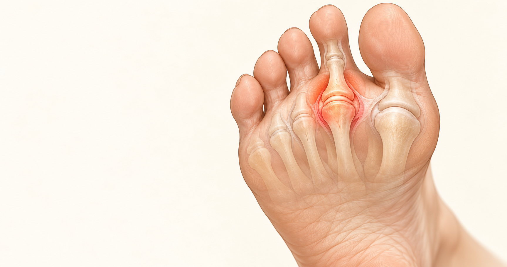
Metatarsalgia caused by capsulitis is pain in the ball of the foot arising from inflammation around one of the metatarsophalangeal joints — the joints where the toes meet the foot. The second metatarsophalangeal joint is particularly vulnerable. Repeated excessive loading can irritate the joint capsule and surrounding tissues, producing pain, tenderness and sometimes swelling beneath the base of the toe. Persistent overload may eventually contribute to weakening of the structures that stabilise the joint, including the plantar plate. Treatment therefore needs to address not only inflammation, but also why excessive pressure is reaching that particular joint.
Metatarsalgia is not a single disease. It is a broad term describing pain around the metatarsal heads and ball of the foot. Capsulitis is one possible cause, alongside plantar plate injury, Morton’s neuroma, stress injury, joint degeneration, Freiberg disease and several other conditions.
Metatarsalgia describes where the foot hurts, not necessarily what is wrong
Patients commonly use the word metatarsalgia as though it were a specific diagnosis. Clinically, it is better understood as a description of forefoot pain.
The metatarsals are the five long bones extending through the forefoot. At their ends are the metatarsal heads, which form joints with the bases of the toes. These are called the metatarsophalangeal joints, or MTP joints. A considerable amount of force passes through this region during walking and running, particularly as the heel rises and the body moves forward over the toes.
Pain underneath this area may therefore come from several different structures. One important source is the MTP joint itself. When the tissues forming and surrounding that joint become repeatedly irritated, an inflammatory condition commonly described as capsulitis can develop.
Capsulitis occurs around the joint at the base of the toe
A joint is not simply two bones touching one another. The metatarsal head and the base of the proximal phalanx are surrounded by a complex system of soft tissues that keep the toe aligned while still allowing it to move. The joint capsule forms a fibrous sleeve around the joint. Its inner lining includes synovial tissue, which helps create the lubricated environment required for normal joint movement.
Around the lesser MTP joints there are also collateral ligaments at the sides and an important fibrocartilaginous structure underneath called the plantar plate. The plantar plate helps prevent the toe from translating excessively upwards over the metatarsal head and is an important stabiliser of the joint. When a joint is repeatedly subjected to more mechanical stress than its tissues are comfortably tolerating, the tissues around it can become irritated and inflamed.
This is the clinical situation commonly described as MTP joint capsulitis. Synovitis may occur at the same time, which is why terminology surrounding early lesser MTP joint problems can sometimes overlap in medical literature. For the patient, however, the important point is simpler: the pain is coming from an overloaded toe joint rather than merely from the skin or metatarsal bone underneath it.
The second toe joint is particularly vulnerable
Capsulitis can occur at several of the lesser metatarsophalangeal joints, but the second MTP joint is particularly important. The second metatarsal forms part of a relatively stable central portion of the foot. During forward progression, substantial loading can pass beneath the second and third metatarsal region.
If the normal distribution of force across the forefoot changes, the second metatarsal head may receive disproportionately high loading. This helps explain why patients frequently develop pain directly underneath the joint at the base of the second toe.
It also explains why simply describing the problem as “pain under the second metatarsal” does not reveal the cause. The bone, joint capsule, plantar plate, adjacent nerves and surrounding soft tissues are all located within a remarkably small anatomical area. Accurate diagnosis matters.
Repeated overload is usually more important than a single painful step
Capsulitis often develops gradually. A person may initially notice mild discomfort underneath a toe after a long walk, run or prolonged period of standing. It settles, returns again after activity, and eventually starts appearing earlier during exercise. At this stage there may be no obvious toe deformity. The problem is often one of repeated mechanical loading.
Every step exposes the affected joint to force. If the forces reaching that joint repeatedly exceed what its tissues can comfortably tolerate, inflammation can persist. This does not necessarily mean the patient is walking or running “incorrectly”. Human feet vary considerably in shape, mobility, bone alignment and load distribution. The important issue is how that particular foot is distributing force.
Forefoot loading can change for several reasons
A painful second MTP joint often cannot be explained by looking only at the painful spot. The mechanics of the entire forefoot matter. One recognised mechanism is first-ray insufficiency. Normally, the first metatarsal and great toe contribute substantially during the later stages of walking. If the first ray does not accept its expected share of load, more pressure may be transferred towards the lesser metatarsals. This is one reason metatarsalgia can occur alongside hallux valgus and other disorders affecting the first ray.
Metatarsal length can also contribute, although the relationship is more complicated than simply saying that everybody with a relatively long second metatarsal will develop pain. Research investigating second MTP instability suggests that metatarsal length is one potential factor among several rather than an isolated explanation.
Other influences can include foot shape, toe deformity, footwear, repetitive forefoot loading, changes in activity and loss of normal load-sharing elsewhere in the foot. High-heeled footwear deserves particular mention because elevating the heel shifts body weight forwards, increasing demand across the forefoot. Chronic forefoot loading associated with high heels has also been recognised in the literature concerning lesser MTP joint instability.
The result is not necessarily one dramatic injury. It may instead be thousands of relatively ordinary steps repeatedly stressing the same small joint.
Capsulitis usually produces very localised ball-of-foot pain
The pain from MTP capsulitis is often centred directly beneath the base of a toe. Patients may describe tenderness when pressing underneath the joint, discomfort during push-off, aching after prolonged walking or a sensation that the area has become bruised. Swelling can sometimes develop around the joint. Movement of the toe may become uncomfortable, particularly when the toe is pushed upwards and the structures beneath the joint are tensioned.
The second MTP joint is the classic location, but similar symptoms may occur at the third or fourth MTP joints. Some patients alter the way they walk because pushing through the painful forefoot becomes uncomfortable. They may unconsciously transfer weight towards the outer edge of the foot or shorten their stride. That compensation does not necessarily solve the original problem and may create additional symptoms elsewhere.
Capsulitis and plantar plate injury are related but they are not the same diagnosis
This distinction is particularly important. The plantar plate is a strong fibrocartilaginous structure beneath the lesser MTP joint. It helps stabilise the toe and resists excessive upward movement of the proximal phalanx over the metatarsal head. Capsulitis describes inflammation around the joint.
A plantar plate injury describes structural damage or deterioration involving an important stabilising structure beneath that joint. A painful MTP joint therefore does not automatically mean that the plantar plate has torn. However, these conditions can exist along the same mechanical pathway.
Persistent repetitive loading can produce inflammation and, in some cases, progressive attenuation of the stabilising tissues. Chronic synovitis has long been discussed in relation to degeneration of the plantar plate and surrounding ligamentous structures.
Once significant plantar plate insufficiency develops, the joint may become mechanically unstable. The toe can then begin to change position. This distinction also explains why early assessment can be valuable. Pain may appear before an obvious deformity develops.
Toe position provides useful information about the stage of the problem
During early capsulitis, the toe may still look completely normal. As instability progresses, subtler changes can develop. The toe may begin losing firm contact with the ground. A small gap may appear between adjacent toes. Eventually the toe can drift sideways or elevate relative to the neighbouring toes.
Advanced instability can progress to subluxation, dislocation or a crossover-toe deformity. Research on lesser MTP instability describes a clinical spectrum ranging from pain and swelling without deformity through increasing instability and eventual malalignment. This does not mean that every case of capsulitis will eventually develop a crooked toe. It means that persistent pain around an MTP joint deserves attention when accompanied by progressive changes in toe position.
Several conditions can feel remarkably similar
The ball of the foot contains many anatomical structures packed into a small space, so symptoms can overlap considerably. Morton’s neuroma may produce forefoot pain but typically involves irritation of an interdigital nerve rather than inflammation of the MTP joint itself. Burning, tingling or electrical sensations extending towards the toes can make a nerve-related source more likely.
A metatarsal stress reaction or stress fracture involves bone rather than the joint capsule. Freiberg disease involves changes affecting the metatarsal head itself. Intermetatarsal bursitis, degenerative joint disease, inflammatory arthropathy, callus-related pressure and plantar plate pathology can also generate pain in a similar region.
Modern imaging reviews of metatarsalgia emphasise this broad differential diagnosis. This is one reason that treating all metatarsalgia with the same generic insole, cushion or shoe is unlikely to be appropriate. The treatment should follow the diagnosis.
Clinical examination looks beyond the painful spot
Assessment normally begins with establishing exactly where the tenderness originates. A podiatrist may examine the painful joint from above and below, assess joint movement and observe the alignment of the toe. The stability of the MTP joint is also important. A commonly used clinical examination is the drawer test, in which the toe is carefully translated relative to the metatarsal head to assess whether abnormal instability is present.
A strongly positive drawer test is particularly useful when plantar plate insufficiency or established MTP instability is suspected. Research comparing clinical examination with operative findings has found the drawer test to be highly specific for plantar plate abnormality. The remainder of the foot should also be assessed.
The great toe, first ray, metatarsal alignment, lesser toes, ankle movement, footwear and walking pattern can all provide information about why the painful joint is being overloaded. This matters because an inflamed joint is often the location of the symptoms, while altered load distribution elsewhere in the foot may be contributing to the reason those symptoms developed.
Imaging is useful when the diagnosis is uncertain or structural injury is suspected
Not every painful MTP joint requires an MRI. Clinical examination often provides substantial information. When imaging is required, weight-bearing X-rays can show the architecture of the forefoot, metatarsal relationships, joint position and associated deformities. Ultrasound can provide useful dynamic assessment of soft tissues.
MRI provides more detailed soft-tissue imaging and is particularly valuable when plantar plate pathology, bone stress injury or another deeper structural disorder is suspected. A recent imaging review describes MRI as the reference modality for detailed soft-tissue assessment in metatarsalgia while recognising the complementary role of ultrasound and weight-bearing radiographs.
The appropriate imaging therefore depends on what the clinician is trying to confirm or exclude.
Treatment should reduce stress on the joint rather than simply suppress the pain
Capsulitis is inflammatory, but the inflammation often exists because the joint continues to be mechanically irritated. Reducing pain without changing the mechanical environment may therefore provide only temporary improvement.
Early management commonly involves reducing aggravating activity while the irritated tissues settle. Footwear can be modified to reduce stress across the forefoot. Depending on the individual foot, this may involve altering forefoot stiffness, cushioning or the way the shoe allows the foot to roll forwards.
For some patients, temporary stabilisation of the affected toe can also reduce repeated stress across the MTP joint. When forefoot pressure distribution is contributing, an appropriately designed orthotic or metatarsal offloading modification may be useful.
Research has demonstrated that metatarsal pads can reduce pressure beneath metatarsal heads, although their effect depends substantially on their position and on the individual foot. Clinical studies have also reported improvements in pain and function in patients with metatarsalgia using appropriately applied metatarsal padding.
This is important because a metatarsal pad is not simply a cushion placed directly underneath the painful joint. Incorrect positioning can fail to offload the area and may occasionally make pressure worse.
Orthotics are most useful when they address the mechanical reason for overload
An orthotic prescribed for capsulitis should have a purpose. For one patient the priority may be reducing pressure beneath a particular metatarsal head. For another it may involve improving first-ray function. Another patient may need greater stability or a change in the way load travels through the foot during propulsion.
Two people with pain beneath the second MTP joint can therefore require quite different orthotic strategies. This is why the word metatarsalgia alone is not enough to prescribe an orthotic. The underlying biomechanics still need to be established.
Injections require particular care around an unstable joint
Corticosteroid can reduce inflammation, but eliminating inflammation temporarily does not correct mechanical overload. This distinction becomes particularly relevant if the plantar plate and other stabilising structures are already compromised.
A 2026 review of non-surgical treatment for lesser MTP instability notes that steroid injections may reduce inflammation but are also associated with concern regarding further attenuation of the ligamentous structures stabilising the joint. An injection should therefore not substitute for establishing why the joint is painful and whether instability is present.
Running and exercise do not necessarily need to disappear permanently
Active people frequently develop forefoot symptoms and understandably worry that they will need to stop running altogether. Capsulitis is fundamentally a problem of load versus tissue tolerance. During an irritated phase, continuing to expose the joint to the same volume and magnitude of loading may prevent it from settling. Temporary modification can therefore be useful.
As symptoms improve and joint stability allows, loading can usually be reintroduced progressively. The objective is not simply rest. It is to create an environment in which the joint is exposed to an amount of load that the tissues can tolerate while the factors responsible for excessive local stress are addressed.
Persistent capsulitis should not be ignored when the toe begins to change position
Simple forefoot soreness after an unusually long day is common. Persistent pain underneath the same toe joint is different. Assessment becomes particularly important when pain repeatedly returns, swelling develops, the toe begins lifting away from the ground, the gap between the toes changes, the toe starts drifting sideways or everyday walking becomes increasingly uncomfortable.
These findings raise concern that the problem may have moved beyond temporary inflammation towards mechanical instability. Once significant deformity develops, treatment becomes considerably more complicated.
The central issue is pressure distribution
Capsulitis illustrates an important principle in foot biomechanics. The site that hurts is not always the place where the entire problem begins. The inflamed second MTP joint may simply be the structure receiving more force than it can tolerate. The cause may involve the relationship between the metatarsals, reduced contribution from the first ray, toe alignment, footwear, repetitive loading or several factors acting together. This is why effective treatment requires more than identifying inflammation.
It requires understanding why that joint became overloaded in the first place. At Family Podiatry Centre, the clinical objective when assessing metatarsalgia is therefore to identify the structure producing the pain, determine whether the MTP joint remains stable and assess the mechanical factors influencing load across the forefoot.
For patients with capsulitis, reducing irritation is important. Reducing the reason for the irritation is usually even more important.
Key takeaway
Metatarsalgia caused by capsulitis occurs when one of the joints at the base of the toes becomes irritated and inflamed, commonly because of repetitive mechanical overload. The second MTP joint is particularly susceptible. Early capsulitis may cause pain and swelling without visible deformity, while persistent overload can sometimes coexist with or progress towards plantar plate insufficiency and MTP joint instability. Because several other conditions produce very similar ball-of-foot pain, accurate diagnosis and assessment of forefoot mechanics are important before deciding on treatment.
Book An Appointment
Disclaimer: The word "treatment" in this article refers to the care and management of a patient’s health to prevent, cure, or improve a condition. Treatment results vary and do not necessarily indicate a cure. This article is for informational and educational purposes only and does not constitute medical advice.
About the Author
Mark B. Reyneker, BTech (Podiatry), MSc (Palaeontology) is a podiatrist and Founder & Clinical Director of Family Podiatry Centre, with more than 25 years of clinical experience across South Africa, Malaysia and Singapore. His clinical interests include foot and lower-limb pain, gait and biomechanics, sports-related foot conditions, orthotic therapy and footwear.
Alongside his clinical practice, Mark conducts research into human gait and foot biomechanics. His MSc research at the University of the Witwatersrand investigated human propulsion and the structural properties of the metatarsals. He is also the inventor of
A Foot Orthotic, an orthotic technology developed through an international patent family.
References
- Ayoola, A.S. and Jastifer, J.R. (2024). Metatarsophalangeal Joint Instability: Anatomy and Physiopathology. Foot and Ankle Clinics, 29(4), 681–690. DOI: 10.1016/j.fcl.2023.12.006.
Available article: https://pubmed.ncbi.nlm.nih.gov/39448180/
- Kinter, C.W. and Hodgkins, C.W. (2020). Lesser Metatarsophalangeal Instability: Diagnosis and Conservative Management of a Common Cause of Metatarsalgia. Sports Health, 12(4), 390–394. DOI: 10.1177/1941738120904944.
Available article: https://www.researchgate.net/profile/Christopher-Kinter/publication/340290222_Lesser_Metatarsophalangeal_Instability_Diagnosis_and_Conservative_Management_of_a_Common_Cause_of_Metatarsalgia/links/5e9cfeb4a6fdcca7892843d5/Lesser-Metatarsophalangeal-Instability-Diagnosis-and-Conservative-Management-of-a-Common-Cause-of-Metatarsalgia.pdf
- Maas, N.M.G., van der Grinten, M., Bramer, W.M. and Kleinrensink, G.J. (2016). Metatarsophalangeal joint stability: a systematic review on the plantar plate of the lesser toes. Journal of Foot and Ankle Research, 9. DOI: 10.1186/s13047-016-0165-2.
Available article: https://link.springer.com/article/10.1186/s13047-016-0165-2
- Männikkö, K. and Sahlman, J. (2017). The Effect of Metatarsal Padding on Pain and Functional Ability in Metatarsalgia. Scandinavian Journal of Surgery, 106(4), 332–337. DOI: 10.1177/1457496916683090.
Available article: https://journals.sagepub.com/doi/full/10.1177/1457496916683090
- Palka, O., Guillin, R., Lecigne, R. and Combes, D. (2025). Radiological approach to metatarsalgia in current practice: an educational review. Insights into Imaging, 16, 94. DOI: 10.1186/s13244-025-01945-3.
Available article: https://link.springer.com/article/10.1186/s13244-025-01945-3
- Yoon, T.H. et al. (2019). Forefoot disorders and conservative treatment. Journal of the Korean Orthopaedic Association / review article available through PubMed Central. Open-access review
Available article: https://www.researchgate.net/profile/Min-Chang-3/publication/333091393_Forefoot_disorders_and_conservative_treatment/links/5cdcb395299bf14d959c47dd/Forefoot-disorders-and-conservative-treatment.pdf