Achilles Tendinopathy: When Ankle Stiffness, Calf Tightness or Heel Pain May Be Coming from the Achilles



10 October 2026
By Mark Reyneker, BTech (Podiatry), MSc (Palaeontology), Registered Podiatrist, Foot & Gait Specialist | Founder & Clinical Director, Family Podiatry Centre
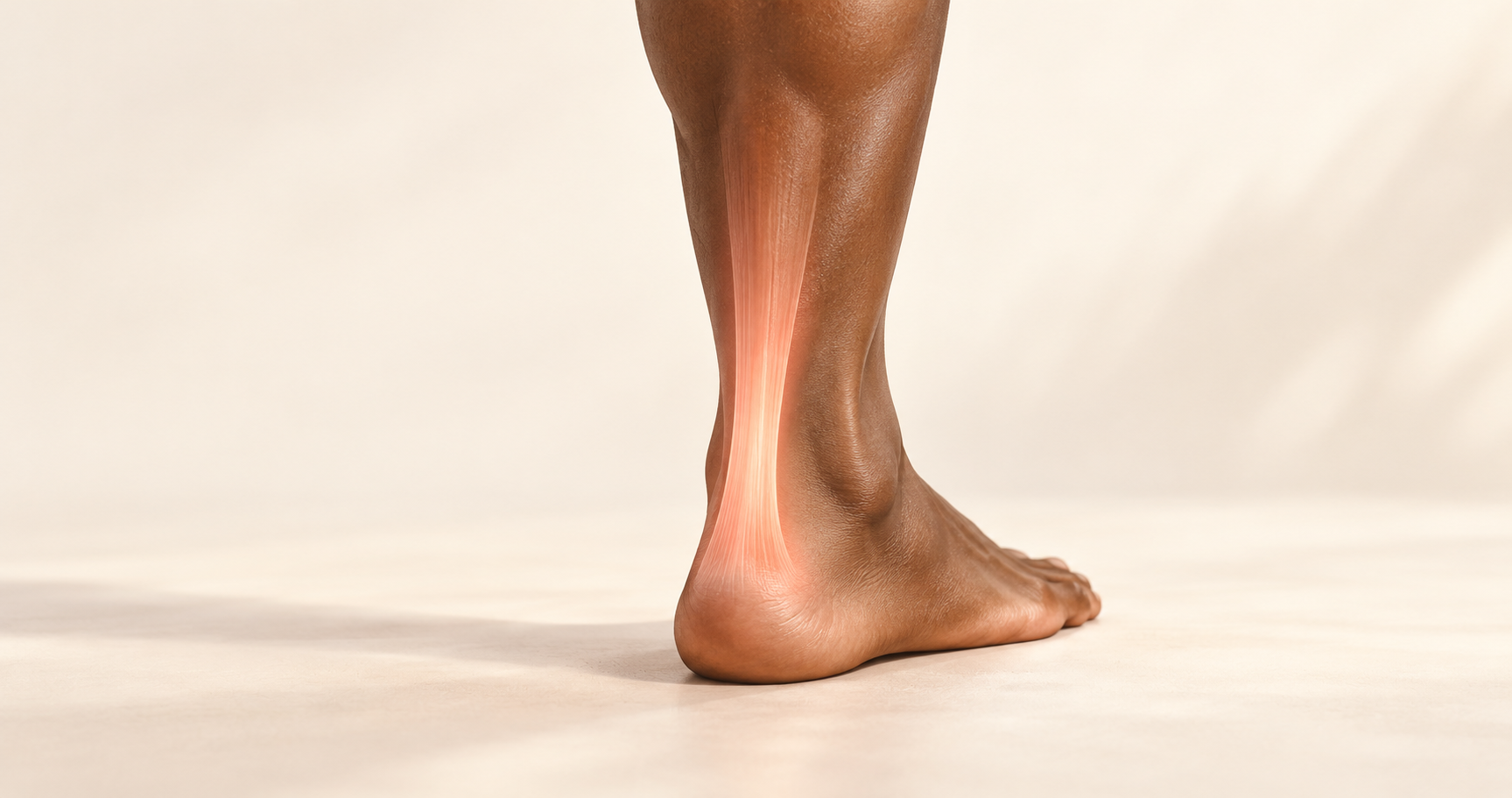
The important point is that Achilles tendinopathy is usually multifactorial. The tendon, calf muscles, ankle movement, foot mechanics, training load and general tendon health all need to be considered rather than attributing the condition to one foot type or movement.
Achilles tendinopathy does not always begin with obvious Achilles pain
- a stiff ankle;
- tightness around the back of the ankle;
- calf tightness;
- recurrent calf cramping;
- stiffness when first standing after sitting;
- a feeling that the foot does not move properly;
- discomfort around the ankle rather than a clearly identified tendon;
- plantar heel pain;
- reduced power when walking uphill or climbing stairs;
- difficulty running normally;
- a feeling that one leg is “tighter” than the other.
Someone experiencing “tight ankle when walking”, “calf tightness when running”, “ankle stiff in the morning” or “heel and ankle pain” may never search for Achilles tendinopathy. Yet those symptoms may justify assessing the Achilles–calf–ankle complex.
Pain is important, but it is not the only symptom
A patient may not identify the Achilles tendon as the painful structure
- pain associated with tendon loading;
- the location of that pain;
- tenderness during examination;
- stiffness;
- functional impairment.
Achilles tendon abnormalities can exist without Achilles pain
The Achilles tendon is much more than a cord connecting the calf to the heel
The major contributors are the: gastrocnemius, which crosses both the knee and ankle; and the soleus, which lies deeper and crosses the ankle. Their tendon fibres combine to form the Achilles tendon before inserting into the posterior calcaneus. This arrangement means that the Achilles forms the mechanical connection between powerful calf muscles and the foot.
The Achilles tendon stores energy when we walk and run
Tendinopathy is not quite the same thing as tendonitis
“Tendonitis” implies that inflammation is the primary pathological process. Modern understanding is more complicated. Chronic tendinopathy involves changes in tendon structure, collagen organisation, cellular activity and tendon matrix rather than simply persistent conventional inflammation.
For this reason, Achilles tendinopathy is generally the preferred clinical term. This does not mean inflammatory processes never occur. It means that chronic tendon disease cannot adequately be explained as inflammation alone.
There are two important locations of Achilles tendinopathy
Ankle stiffness can be one of the most noticeable complaints
Ankle equinus is not actually a type of foot movement
Pronation and supination are movements.
Equinus describes restriction of ankle dorsiflexion.
A person with ankle equinus cannot move the shin sufficiently forwards over the foot without compensating somewhere else.
One common reason is gastrocnemius tightness. Because the gastrocnemius crosses both the knee and ankle, its effect on ankle dorsiflexion changes according to knee position. This relationship forms the basis of the Silfverskiöld test, which helps clinicians determine whether limited dorsiflexion is substantially influenced by the gastrocnemius. Equinus therefore deserves separate consideration from pronation and supination.
Pronation is the foot movement most commonly associated with Achilles tendinopathy
An earlier systematic review found increased rearfoot eversion range of motion in runners with Achilles tendinopathy. A later systematic review again identified increased ankle eversion and differences in the timing of maximum pronation. More recent research has described an overall tendency towards what investigators called a pattern of medial collapse, although the authors emphasised that study quality remains limited and biomechanical findings are not completely consistent.
Overpronation should not become the universal explanation
Supination can also alter Achilles loading
Reduced ankle dorsiflexion may be as clinically important as foot posture
Something has to compensate. The foot may pronate more. The heel may lift earlier. The leg may rotate differently. The stride may change. The person may shorten the step. The knee or hip may compensate.
The evidence for equinus as a cause is still mixed
One addresses whether limited dorsiflexion predicts the development of the condition. The other demonstrates that reduced dorsiflexion is commonly found once the condition exists. We therefore should not claim: “Equinus causes Achilles tendinopathy.”A more accurate statement is: reduced ankle dorsiflexion is commonly relevant in Achilles tendinopathy and should be assessed, but it is only one component of a multifactorial condition.
Calf tightness and calf cramping deserve attention
Some notice that one calf cramps more easily during running or at night. Calf cramping is not a specific diagnostic sign of Achilles tendinopathy. Many neurological, metabolic, circulatory and muscular factors can produce cramps. However, Achilles tendinopathy is associated with measurable deficits in the plantar-flexor system.
Foot stiffness can be part of the same mechanical picture
- how long the heel remains on the floor;
- where pressure travels under the foot;
- how quickly they move over the forefoot;
- how much the rearfoot everts;
- how strongly the calf generates propulsion.
Plantar heel pain may coexist with Achilles and calf problems
A systematic review examining plantar fasciopathy described the close anatomical and mechanical relationship between the gastrocnemius–Achilles complex and the plantar fascia. Increased Achilles tension and reduced dorsiflexion may increase loading through the plantar fascia during weightbearing.
This does not mean plantar heel pain is a symptom that proves Achilles tendinopathy. Plantar fasciopathy, heel-pad disorders, nerve entrapments, stress injury and other conditions remain important causes of plantar heel pain. But when plantar heel pain occurs alongside: calf tightness + limited ankle dorsiflexion + Achilles stiffness the entire calf–ankle–foot system deserves examination rather than treating the painful heel in isolation.
The Achilles can be abnormal before it hurts
This supports an important concept: pain is one stage of the clinical presentation, not necessarily the beginning of the biological process. A runner may first notice reduced spring. Another notices persistent calf fatigue. Another feels stiff every morning. Another experiences obvious Achilles pain.
Morning stiffness is particularly characteristic
Some patients first notice loss of performance
The Achilles tendon and plantar-flexor muscles are fundamental to propulsion. Recent systematic-review evidence found people with Achilles tendinopathy performed approximately 10 fewer heel raises than asymptomatic controls in pooled testing. Reduced performance therefore deserves attention even when pain is relatively mild.
Achilles tendinopathy is not just a running injury
Age, metabolic health, cholesterol, diabetes, medications, previous injury, tendon capacity and sudden changes in activity can all influence tendon behaviour. Fluoroquinolone antibiotics are a particularly recognised medication-related risk because of their association with Achilles tendon disorders and rupture. The clinical assessment therefore needs to extend beyond simply asking how somebody runs.
The entire lower limb participates in Achilles loading
the foot;
- ankle;
- calf;
- knee;
- hip;
- muscle activation;
- plantar-flexor strength;
- hip strength;
- running mechanics.
Pronation versus supination versus equinus: the practical conclusion
Some of the strongest repeated dynamic observations involve increased rearfoot eversion and altered pronation timing. Pronation is therefore the foot movement most commonly associated with Achilles tendinopathy in running-biomechanics research. It is not present in every patient and should not automatically be considered the cause.
Supination is less commonly presented as the classic Achilles tendinopathy pattern. However, lateral foot roll-over and relatively rigid mechanics may also alter tendon loading. A supinated foot should therefore not be dismissed.
Equinus is not a movement pattern. It describes restricted ankle dorsiflexion. Reduced dorsiflexion is commonly found clinically in Achilles tendinopathy and may influence loading and compensatory foot movement. However, research has not conclusively established equinus as a universal cause.
The location of symptoms still matters
Pain anterior to the Achilles may suggest posterior ankle impingement. Pain around the side of the tendon may involve neighbouring tendons or nerves. Plantar heel pain requires a different differential diagnosis. Deep calf pain and swelling may occasionally indicate something requiring urgent medical assessment rather than a tendon disorder.
An international Delphi consensus published in 2025 identified several important alternative diagnoses around the Achilles region, including partial tendon tear, posterior ankle impingement, plantaris pathology, other flexor-tendon disorders, paratendinopathy, nerve pathology, bursitis, calcaneal stress injury and ankle or subtalar pathology.
Imaging is useful when it answers a clinical question
Treatment should follow the diagnosis and the mechanical findings
Persistent ankle stiffness and calf symptoms deserve investigation
- whether the Achilles tendon is actually involved;
- whether the condition is insertional or midportion;
- whether another structure is producing the symptoms;
- how much ankle dorsiflexion is available;
- whether gastrocnemius or soleus restriction is present;
- how the rearfoot moves;
- whether the patient predominantly pronates or supinates during gait;
- how pressure moves through the foot;
- how effectively the calf produces propulsion;
- and what activity or loading factors may have contributed to the problem.
Key takeaway
Disclaimer: The word "treatment" in this article refers to the care and management of a patient’s health to prevent, cure, or improve a condition. Treatment results vary and do not necessarily indicate a cure. This article is for informational and educational purposes only and does not constitute medical advice.
About the Author
Mark B. Reyneker, BTech (Podiatry), MSc (Palaeontology) is a podiatrist and Founder & Clinical Director of Family Podiatry Centre, with more than 25 years of clinical experience across South Africa, Malaysia and Singapore. His clinical interests include foot and lower-limb pain, gait and biomechanics, sports-related foot conditions, orthotic therapy and footwear.
Alongside his clinical practice, Mark conducts research into human gait and foot biomechanics. His MSc research at the University of the Witwatersrand investigated human propulsion and the structural properties of the metatarsals. He is also the inventor of A Foot Orthotic, an orthotic technology developed through an international patent family.
References
- Docking, S.I., Hart, H.F., Rio, E., Hannington, M.C., Cook, J.L. and Culvenor, A.G. (2021). Explaining variability in the prevalence of Achilles tendon abnormalities: a systematic review with meta-analysis of imaging studies in asymptomatic individuals. Journal of Orthopaedic & Sports Physical Therapy, 51(5), pp.232–252. DOI: 10.2519/jospt.2021.9970.
Available article: https://pubmed.ncbi.nlm.nih.gov/33779214/ - Johannsen, F.E., Rydahl, J.P., Jacobsen, A.S., Brahe, C.C.H. and Magnusson, P.S. (2024). Foot Posture and Ankle Dorsiflexion as Risk Factors for Developing Achilles Tendinopathy and Plantar Fasciitis: A Case-Control Study. Foot & Ankle International, 45(12), pp.1380–1389. DOI: 10.1177/10711007241281289.
Available article: https://europepmc.org/article/med/39422991 - Martin, R.L. et al. (2024). Achilles Pain, Stiffness, and Muscle Power Deficits: Midportion Achilles Tendinopathy Revision 2024. Journal of Orthopaedic & Sports Physical Therapy. DOI: 10.2519/jospt.2024.0302.
Available article: https://www.orthopt.org/uploads/content_files/files/chimenti_et_al_2024_achilles_pain_stiffness_and_muscle_power_
deficits_midportion_achilles_tendinopathy.pdf - McAuliffe, S. et al. (2025). Achilles tendinopathy physical impairments evaluated through clinician-friendly measures: a systematic review with meta-analysis and GRADE recommendations.
Available article: https://www.sciencedirect.com/science/article/abs/pii/S1413355525000413 - Munteanu, S.E. and Barton, C.J. (2011). Lower limb biomechanics during running in individuals with Achilles tendinopathy: a systematic review. Journal of Foot and Ankle Research, 4, 15. DOI: 10.1186/1757-1146-4-15.
Available article: https://onlinelibrary.wiley.com/doi/full/10.1186/1757-1146-4-15 - Ogbonmwan, I., Kumar, B.D. and Paton, B. (2018). New lower-limb gait biomechanical characteristics in individuals with Achilles tendinopathy: a systematic review update. Gait & Posture, 62. DOI: 10.1016/j.gaitpost.2018.03.010.
Available article: https://www.sciencedirect.com/science/article/abs/pii/S0966636218301425 - Quarmby, A., Mönnig, J., Mugele, H., Henschke, J., Kim, M., Cassel, M. and Engel, T. (2023). Biomechanics and lower limb function are altered in athletes and runners with Achilles tendinopathy compared with healthy controls: a systematic review. Frontiers in Sports and Active Living, 4, 1012471. DOI: 10.3389/fspor.2022.1012471.
Available article: https://www.frontiersin.org/journals/sports-and-active-living/articles/10.3389/fspor.2022.1012471/full
Frequently Asked Questions
Early symptoms can include morning ankle stiffness, tightness around the back of the ankle, calf tightness, reduced push-off, discomfort after exercise and a gradual decline in running or walking performance. Some patients do not initially describe obvious Achilles tendon pain.
Yes. Stiffness, particularly after rest or first thing in the morning, is a common feature of Achilles tendinopathy. Some people notice tightness or restricted ankle movement before pain becomes their main complaint.
Achilles disorders can be associated with altered calf function, fatigue and reduced plantar-flexor endurance. Some patients describe calf tightness or cramping, although cramps are not specific to Achilles tendinopathy and can have many other causes.
Increased rearfoot eversion and altered pronation timing have been reported in some people with Achilles tendinopathy, particularly runners. However, overpronation is not a universal cause, and many people with Achilles tendinopathy do not have an excessively pronated foot.
Pronation, particularly increased rearfoot eversion, is more consistently reported in Achilles tendinopathy research than supination. However, supinated or lateral loading patterns can also alter Achilles loading, so foot mechanics need to be assessed individually.
Reduced ankle dorsiflexion, sometimes described clinically as ankle equinus when sufficiently restricted, is commonly found in people with Achilles tendinopathy. It may alter how the foot and lower limb compensate during walking and running, although it should not be considered a universal cause.
Achilles tendinopathy and plantar heel pain are separate conditions, but they can coexist. Restricted ankle dorsiflexion and calf tightness may influence both the Achilles tendon and plantar fascia, so patients with heel pain and ankle stiffness may benefit from assessment of the entire calf–ankle–foot system.
Yes. Ultrasound and MRI studies frequently identify structural Achilles tendon abnormalities in people who have no symptoms. An abnormal scan does not automatically mean a person has symptomatic Achilles tendinopathy.
Midportion Achilles tendinopathy usually affects the tendon several centimetres above the heel. Insertional Achilles tendinopathy occurs where the tendon attaches directly to the heel bone. The two conditions involve different mechanical environments and may require different management strategies.
Assessment is advisable when stiffness, calf tightness, reduced push-off or posterior ankle discomfort becomes persistent, repeatedly returns, affects activity or is accompanied by local Achilles tenderness. Similar symptoms can also arise from other ankle, tendon, nerve or muscular conditions.